, Maria Custódia Machado Ribeiro2 and Bruno Beber Machado3
(1)
Hospital da Criança de Brasília – Jose Alencar, Clínica Vila Rica, Brazília, Brazil
(2)
Hospital da Criança de Brasília – Jose Alencar, Hospital de Base do Distrito Federal, Brasília, Brazil
(3)
Clínica Radiológica Med Imagem, Unimed Sul Capixaba, Santa Casa de Misericordia de, Cachoeiro de Itapemirim, Brazil
Abstract
The juvenile spondyloarthropathies comprise a group of conditions characterized by inflammation of the sacroiliac joints and of the spine, association with HLA-B27 antigen, and disease onset before age 16. This group encompasses juvenile ankylosing spondylitis, juvenile psoriatic arthritis, reactive arthritis, arthritis associated with inflammatory bowel disease, and the undifferentiated forms of spondyloarthropathy. In addition to the axial skeleton, the juvenile spondyloarthropathies also affect the peripheral joints, mostly in the lower extremities, and prominent enthesopathy is often present. Peripheral arthritis usually predominates during the initial stage and sacroiliitis/spondylitis are late findings, so that children with early-stage juvenile spondyloarthropathies may be incorrectly diagnosed as having classic juvenile idiopathic arthritis. Classic radiographic criteria employed for adult-type disease are fairly insensitive for early diagnosis of juvenile spondyloarthropathies. Therefore, imaging methods such as ultrasonography (US) and magnetic resonance imaging (MRI), which are more sensitive, have become more and more important, given that early diagnosis and timely introduction of treatment are paramount for improved outcome and better quality of life. Pediatric collagen vascular disorders are multisystemic autoimmune diseases of unknown etiology, which are similar in many ways to their adult counterparts. Nonetheless, peculiarities in the presentation and in the evolution of the juvenile forms of collagen diseases make them unique, thus deserving separate study. Even though osteoarticular manifestations are not the more relevant in most patients, some imaging findings are quite typical, and it is important for the radiologist to be acquainted with them. The following paragraphs will discuss the major juvenile spondyloarthropathies and the juvenile forms of dermatomyositis, systemic lupus erythematous, scleroderma, and mixed connective tissue disease.
4.1 Introduction
The juvenile spondyloarthropathies comprise a group of conditions characterized by inflammation of the sacroiliac joints and of the spine, association with HLA-B27 antigen, and disease onset before age 16. This group encompasses juvenile ankylosing spondylitis, juvenile psoriatic arthritis, reactive arthritis, arthritis associated with inflammatory bowel disease, and the undifferentiated forms of spondyloarthropathy. In addition to the axial skeleton, the juvenile spondyloarthropathies also affect the peripheral joints, mostly in the lower extremities, and prominent enthesopathy is often present. Peripheral arthritis usually predominates during the initial stage and sacroiliitis/spondylitis are late findings, so that children with early-stage juvenile spondyloarthropathies may be incorrectly diagnosed as having classic juvenile idiopathic arthritis. Classic radiographic criteria employed for adult-type disease are fairly insensitive for early diagnosis of juvenile spondyloarthropathies. Therefore, imaging methods such as ultrasonography (US) and magnetic resonance imaging (MRI), which are more sensitive, have become more and more important, given that early diagnosis and timely introduction of treatment are paramount for improved outcome and better quality of life.
Pediatric collagen vascular disorders are multisystemic autoimmune diseases of unknown etiology, which are similar in many ways to their adult counterparts. Nonetheless, peculiarities in the presentation and in the evolution of the juvenile forms of collagen diseases make them unique, thus deserving separate study. Even though osteoarticular manifestations are not the more relevant in most patients, some imaging findings are quite typical, and it is important for the radiologist to be acquainted with them. The following paragraphs will discuss the major juvenile spondyloarthropathies and the juvenile forms of dermatomyositis, systemic lupus erythematous, scleroderma, and mixed connective tissue disease.
4.2 Juvenile Spondyloarthropathies
4.2.1 Juvenile Ankylosing Spondylitis
Juvenile ankylosing spondylitis (JAS) is more frequent in males, and disease onset usually occurs between the end of childhood and the first years of adolescence. Arthritis and enthesitis (inflammation involving the sites of insertion of tendons, ligaments, and joint capsules) are the more important articular manifestations in JAS. In contradistinction to the adult form of ankylosing spondylitis, in which axial involvement predominates from the start, in JAS, years (or even decades) may elapse before spondylitis and sacroiliitis become evident.
Arthritis is more common in large joints – mainly in the lower extremities – and the peripheral small joints are involved less often. If compared to that found in adult-type ankylosing spondylitis, articular disease in JAS usually follows a more severe course, with unrelenting arthritis, disability, and occasional need for joint replacement (Figs. 4.1 and 4.2). Radiographic findings related to peripheral arthritis include progressive narrowing of joint space and bone erosions of late appearance (Fig. 4.1). MRI is the method of choice for early diagnosis, demonstrating joint effusion, synovial thickening, and inflammation of the soft tissues and of the bones (Fig. 4.1), even in the pre-erosive stage. US is also able to demonstrate joint effusion, synovitis, and soft-tissue swelling as soon as they appear. Enthesitis is frequently found in the insertions of the plantar fascia, of the knee extensor mechanism, and of the Achilles tendon. Enthesitis-related radiographic findings include periosteal reaction, bone spurs, and enthesophytes, as well as sclerosis and lucent areas in the adjacent bone. Periostitis is more common in the hands, while enthesophytes are more frequently seen in the tarsal bones (Fig. 4.1). The affected tendon appears thickened and heterogeneous on US and on MRI, with increased perfusion on Doppler US and post-gadolinium enhancement. Cortical erosions and bone marrow edema pattern are also present, as well as soft-tissue swelling and fluid distention of adjacent bursae.
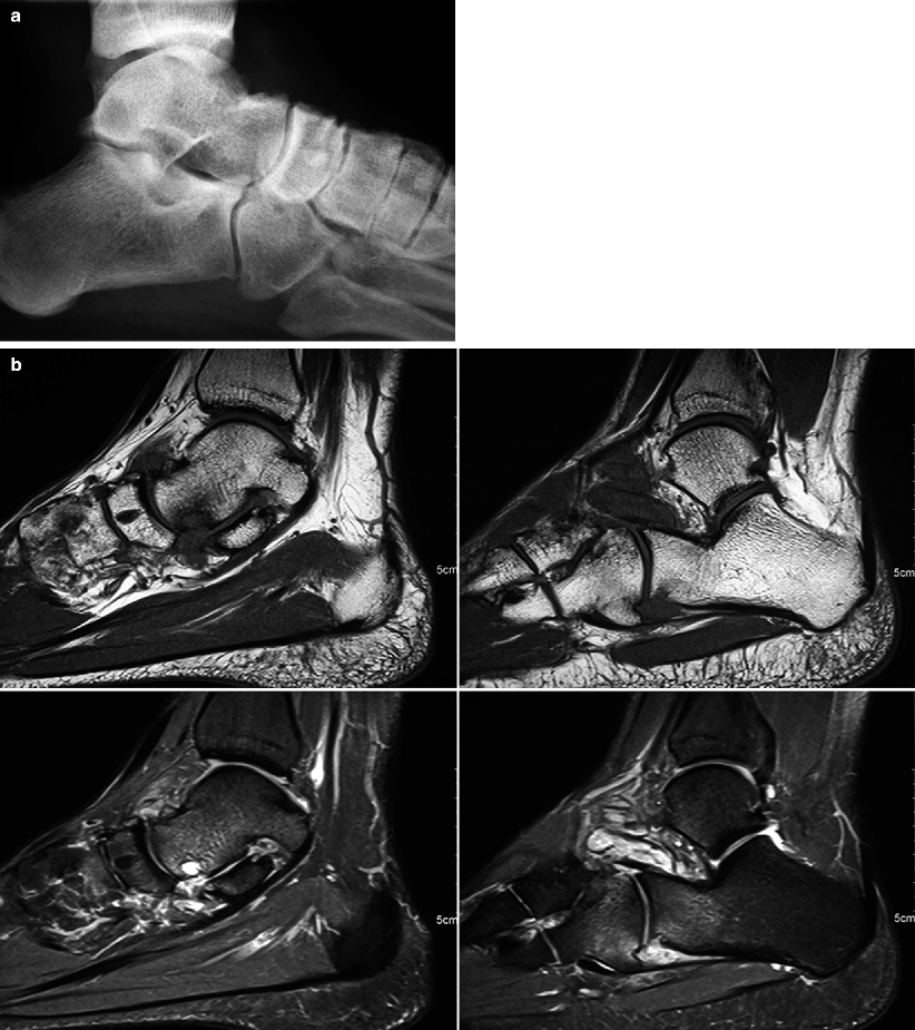
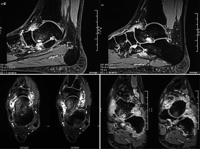
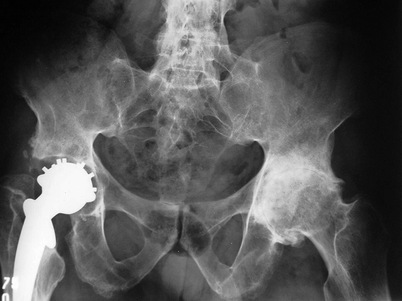


Fig. 4.1
An 18-year-old male with arthritis of the left midfoot and hindfoot since 3 years before. Lateral view (a) of the ankle demonstrates dorsal enthesophytosis in the midfoot and subcortical lucencies in the lowermost portion of the cuboid bone. In (b), sagittal T1-WI (upper row) and fat sat PD-WI (lower row) of the same ankle reveal arthritic changes, with joint effusion, synovial thickening, and bone marrow edema, as well as erosions in the talus, in the calcaneus, and in the cuboid. Post-gadolinium images (c) disclose enhancement of the thickened synovium. Tarsitis is a peculiar manifestation of juvenile spondyloarthritides, representing a combination of inflammation and bone proliferation that often leads to ankylosis

Fig. 4.2
Pelvic radiograph of a 27-year-old patient with JAS since late adolescence. There is severe osteoarthritis of the left hip (secondary to chronic arthritis) and right-sided sacroiliitis with pseudo-widening of the joint space, as well as ankylosis of the left sacroiliac joint and of the lower lumbar facet joints. Total replacement of the right hip joint is also seen, with a wide lucent zone surrounding the acetabular component of the prosthesis, indicative of loosening
Sacroiliitis in JAS is prototypical, very similar to that found in adult-type disease. When positive, radiographs show loss of definition of the articular surfaces, bone erosions, and subchondral sclerosis in the sacroiliac joints, which are most often bilateral. As the disease advances, there is gradual narrowing of the joint spaces, and ankylosis may be found in late-stage JAS. Pseudo-widening of the joint space is a peculiar finding, caused by confluence of subchondral erosions (Figs. 4.2 and 4.3). However, radiographs are insensitive for the assessment of the sacroiliac joints, largely because of their complex anatomy; furthermore, the classic radiographic findings are only found in advanced sacroiliitis (Fig. 4.2), and this imaging method is unable to distinguish active from inactive disease. In spite of the excellent bone assessment provided by computed tomography (CT) (Fig. 4.3), this imaging modality is also unable to yield accurate information about inflammation activity. MRI is the most suitable study for sacroiliac evaluation, as it is able to detect subchondral areas of bone marrow edema that show post-gadolinium enhancement and are indicative of active disease (Fig. 4.4); these areas of osteitis are evident since the early stages of sacroiliitis, way before the development of erosions. Increased signal intensity on T2-weighted images (T2-WI) and post-gadolinium enhancement can also be seen inside the articular space in inflamed sacroiliac joints. Bone erosions are usually small, and large subchondral cysts are relatively rare (Fig. 4.5); subchondral sclerosis appears as areas of low signal intensity in all sequences (Fig. 4.5). Fatty replacement of bone marrow is a late finding, indicative of healing and typically found in chronic sacroiliitis. Despite the high sensitivity of bone scintigraphy, its usefulness is limited in the assessment of sacroiliitis in the juvenile spondyloarthropathies, mostly because of technical issues in the evaluation of the immature sacroiliac joints (see Chap. 1) and due to its inherently low specificity and spatial resolution (Fig. 4.6).
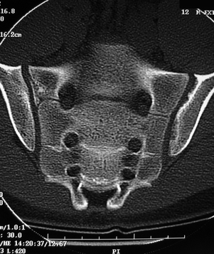
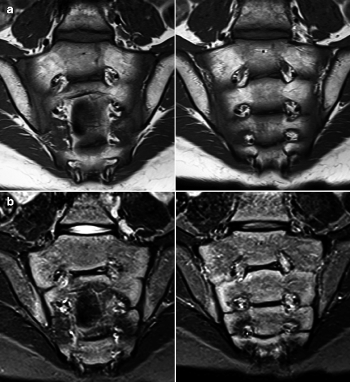
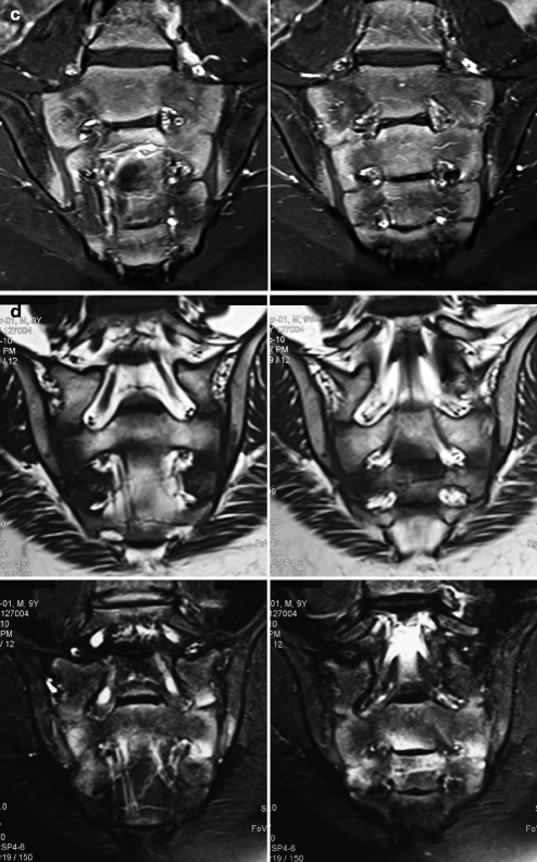
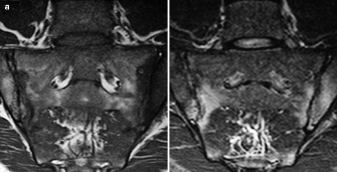
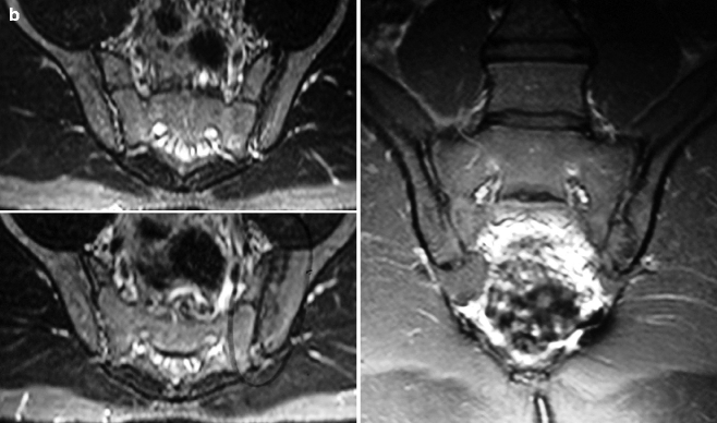
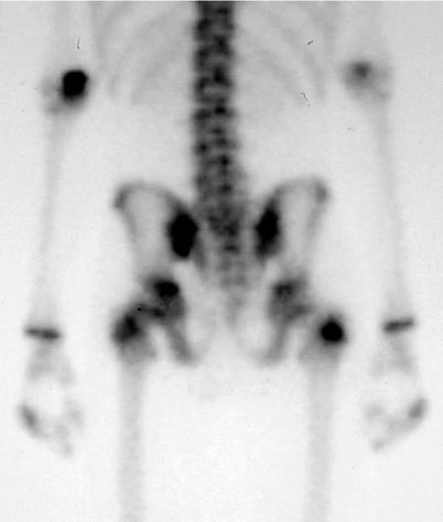

Fig. 4.3
A 12-year-old male with JAS. Coronal CT image shows confluent subchondral erosions in the iliac surface of the left sacroiliac joint, with subtle sclerosis of the adjacent bone. This confluence leads to pseudo-widening of the sacroiliac joint space, similar to that seen in the right sacroiliac joint in the patient of Fig. 4.2


Fig. 4.4
Coronal T1-WI (a), STIR image (b), and post-gadolinium fat sat T1-WI (c) of a 13-year-old child with JAS and pre-erosive sacroiliitis. There is subchondral bone marrow edema pattern along the sacral surfaces and in the lowermost portions of the iliac surfaces of both sacroiliac joints, more prominent at right, with post-contrast enhancement. Joint spaces are preserved and there are no erosions. Similar findings are seen in (d) in a 9-year-old male with undifferentiated spondyloarthropathy (upper row, T1-WI; lower row, fat sat T2-WI)


Fig. 4.5
MRI of the sacroiliac joints of two different patients. In (a), in a 19-year-old male with JAS since age 15, there is bilateral subchondral osteitis, which appears as areas of low signal intensity on T1-WI (left image) and high signal intensity on STIR (right image). There is narrowing of the joint spaces and irregularity of the articular surfaces due to micro-erosions; one dominant bone erosion is seen in the upper half of the left sacral surface. In (b), MRI of a 14-year-old male with JAS and good clinical control shows narrowing of the left sacroiliac joint space and subchondral sclerosis, which appears as hypointense areas on transverse T2-WI (left images) and coronal post-gadolinium fat sat T1-WI (right image). Absence of bone marrow edema or abnormal post-contrast enhancement is indicative of inactive disease

Fig. 4.6
Bone scintigraphy of the same patient of Fig. 4.3, posterior view. There is increased uptake in the lowermost portion of the left sacroiliac joint, indicative of active inflammation. Radiographs and CT are insensitive for assessment of disease activity
Spondylitis usually occurs in late-stage JAS and is rarely found in children, affecting most often the thoracolumbar spine of adults with long-standing disease (Fig. 4.2); sacroiliitis almost always precede vertebral involvement. Involvement of the cervical spine is rare and late, presenting a more benign course if compared to that seen in juvenile idiopathic arthritis. Classic radiographic findings (infrequently seen in skeletally immature patients) include sclerosis of vertebral corners, squaring of the vertebral bodies, syndesmophytosis, and ankylosis of the facet joints.
4.2.2 Juvenile Psoriatic Arthritis
Juvenile psoriatic arthritis (JPA) is a form of seronegative arthritis associated with psoriasis, much less common and more subtle than the adult form of the disease. The typical cutaneous rash is absent in up to half of the patients. This is the only spondyloarthropathy in which females are more frequently affected (up to 2.5:1), with an incidence peak at 11–12 years of age. Diagnosis is based on clinical features, a compatible family history, laboratory tests, and imaging findings.
Arthritis is usually oligoarticular and asymmetric, affecting most often the knees and the small joints of the hands and feet. Radiographs are insensitive for early-stage JPA; the association of periostitis, bone erosions, and enthesitis is very characteristic of JPA, but these are late findings, as well as sacroiliitis/spondylitis. The hands are affected most often and more severely than the feet, and arthritis of the distal interphalangeal joints is typical (Fig. 4.7). Peripheral erosions may lead to the classic “mouse ear” appearance (Fig. 4.8). Arthritis mutilans is a rare and severe form of JPA, presenting imaging findings similar to those of juvenile idiopathic arthritis (Figs. 4.9 and 4.10). Abnormalities commonly found in the adult-type form of the disease, such as acro-osteolysis (resorption of the distal phalanges), whiskering (proliferative bone neoformation in the entheses), and periostitis tend to be less aggressive in JPA. If compared to JAS, there is more extensive involvement of the small joints and less enthesopathy in JPA, features that help to narrow the differential diagnosis. The so-called “pencil-in-cup” deformity, a classic finding of psoriatic arthritis in adults, is uncommonly seen in children (Figs. 4.8 and 4.11).
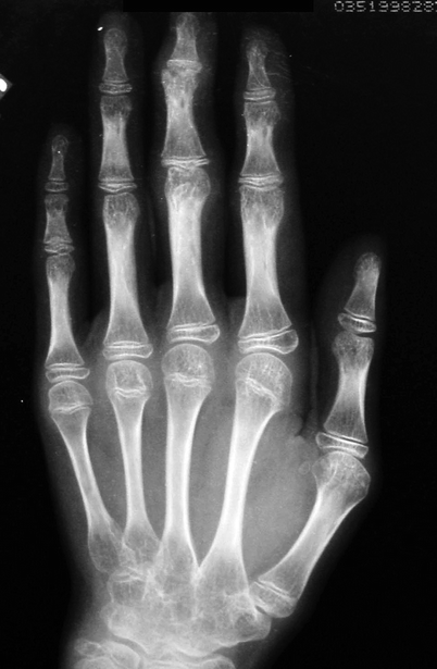
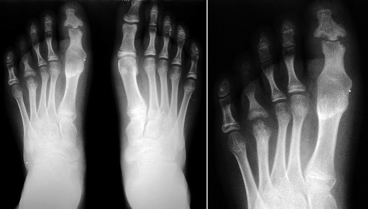

Fig. 4.7
A 12-year-old female with severe JPA. Erosive arthritis can be seen in the interphalangeal joints of the third finger, mainly distal. Solid periosteal reaction along the diaphyses of the proximal phalanges is also present, from the second to the fourth digits. Extensive carpal ankylosis is evident

Fig. 4.8


Radiograph of the feet of a child with JPA (magnification of the right forefoot in the right image). Erosive arthritis is seen in the interphalangeal joint of the right hallux (“mouse ear” appearance), as well as ankylosis of the corresponding metatarsophalangeal joint. Arthritis of the metatarsophalangeal joint of the fourth toe is also evident, with the classic “pencil-in-cup” configuration, characterized by bone remodeling and a saucerlike appearance of the joint surface of the proximal phalanx, on which the severely eroded metatarsal head articulates. Periostitis is seen along the proximal phalanx of the fifth toe at right and along the diaphysis of the fourth metatarsal bone
Stay updated, free articles. Join our Telegram channel

Full access? Get Clinical Tree


