Subtalar Joint Arthroscopy
Keywords
• Subtalar • Arthroscopy • Portals
Diagnostic tests
One of the most beneficial diagnostic tests is a selective block of the subtalar joint. The patient is prepared and injected into the sinus tarsi/subtalar joint with ropivacaine. Marcaine can also be used, but there is concern for chondrocyte toxicity.1 The patient is instructed to resume normal activities with attention to those activities that they could not perform before the block. An intracapsular problem should achieve significant-to-total relief of symptoms from the block.
Diagnostic tests such as computed tomographic scans, bone scans, and magnetic resonance imaging (MRI) can be used but may have limited diagnostic value. Goldberger and Conti2 concluded that arthroscopy of the subtalar joint was “the most accurate method of diagnosing subtalar articular cartilage damage …” Their study compared plain radiographs, MRI, bone scans, and intraoperative findings of subtalar joint arthroscopy. Lee and colleagues3 concluded that MRI was effective for determining cervical ligament tears, alterations of fat within the sinus tarsi, and synovial thickening but was inaccurate in correctly detecting interosseous ligament tears. Oloff and colleagues4 reported that “magnetic resonance imaging was useful in identifying subtalar joint chronic synovitis and/or fibrosis in all 26 patients who were imaged.”
Results
Subtalar joint arthroscopy has high patient satisfaction rates, with minimal complications. Williams and Ferkel5 reported 86% good-to-excellent results in a subgroup of 29 patients, all with demonstrable arthroscopic pathology. Ahn and colleagues6 reported on 115 consecutive patients who underwent arthroscopy. This study was performed on a wide variety of cases, including arthroscopic subtalar arthrodesis, and found a 97% patient satisfaction rate, with no serious complications reported. Frey and colleagues7 reviewed 49 subtalar arthroscopies, with an average follow-up of more than 4 years. Their excellent-to-good results were 94%, with all the patients with 6% poor result eventually having successful subtalar joint arthrodesis. There were 5 reported complications of the 49 procedures. There were no reported complications in the study by Williams and Ferkel5 or Goldberger and Conti.2
Instrumentation
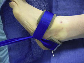
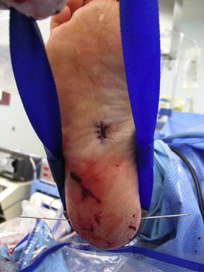
Subtalar joint arthroscopy can be performed with or without distraction. Invasive distraction is rarely used but can be achieved with the use of a monolateral external fixator. The distal arm of the fixator is rotated allowing both pins to enter the calcaneus. Noninvasive distraction can be accomplished by tying Kerlix around the patient’s subtalar joint and around the surgeon’s waist. The surgeon leans back, creating distraction at the joint. There are also several commercial noninvasive distractors on the market. Noninvasive distraction is a 3-part system. It consists of a leg holder, the distraction apparatus that attaches to the operating table, and a heel strap. Because of the nature of the procedure, the skin is wet and the strap from the noninvasive distractor can frequently slip off the back of the heel. Mastisol, placed on the heel, will help prevent the strap from slipping, but if this continues, minimally invasive distraction will prevent the heel strap from slipping. A 0.62 inches Kirschner wire is placed transversely across the calcaneus, and the posterior strap is placed over the wire (Figs. 1 and 2). Wires larger than 0.62 inches can create too much traction. This has produced neuritis specifically at the deep and superficial peroneal nerves.
Procedure
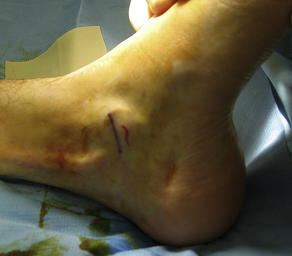
The portals are marked before insufflation to prevent losing the anatomic boundaries once the joint is distended (Fig. 3). The subtalar joint can be approached through several portals: lateral, accessory lateral, medial,8 middle,9 posteromedial, and posterolateral.10 The author’s preferred technique is to use anterolateral and middle portals. These portals are easily developed by palpating into the sinus tarsi, anterior to the fibula, and posterior to the beak of the calcaneus (Figs. 4–6). When a posterior approach is needed, a switch stick is used in the middle portal. This technique is visualized through the anterolateral portal. Once the position of the portal has been determined by the switch stick’s protrusion on the posterolateral aspect of the ankle/subtalar joint, a small incision is made over the protruding switch stick and an arthroscopic cannula is then placed over the switch stick (Figs. 7 and 8). This allows direct access to the posterior aspect of the subtalar joint. A 70° arthroscope is placed into the middle portal for visualization, whereas the posterolateral portal is used as a working portal.


Stay updated, free articles. Join our Telegram channel

Full access? Get Clinical Tree


