Jaundice (Case 25)

Jaundice (Case 25)


Austin Hwang MD and Giancarlo Mercogliano MD, MBA, AGA


Case: A 60-year-old man presents with jaundice, 20-pound weight loss, intermittent nausea, and decreased appetite over the last month. He has a history of hypertension, hyperlipidemia, and diabetes. There is no past surgical history. He takes hydrochlorothiazide, simvastatin, and metformin. His BP, cholesterol, and diabetes are under good control. He has drunk three beers each day and smoked half a pack of cigarettes per day for the last 40 years. He has no abdominal pain, but he has noticed that his stools have become lighter in color and his urine has become tea-colored. He presents in the outpatient office accompanied by his wife and three of his children, who have urged him to seek medical attention.


Differential Diagnosis


Hepatocellular Causes


Extrahepatic/Obstructive Causes


Viral hepatitis


Choledocholithiasis


Alcoholic hepatitis


Cholangitis


Drug-induced hepatitis


Benign stricture


Cirrhosis


Pancreatic adenocarcinoma


Speaking Intelligently


When asked to see an older patient with jaundice, we worry about cancer. A helpful start in patients with this clinical presentation is to decide whether the cause is hepatocellular or obstructive. These two categories serve as a useful framework to think about elevated serum bilirubin levels. Treatment of hepatocellular causes is generally supportive, while treatment of obstructive causes is with endoscopy or surgery. History taking allows me to create a diagnostic hypothesis. Laboratory values and imaging help me to corroborate this hypothesis. Liver function tests (LFTs) are crucial. Ultrasonography evaluates the hepatic parenchyma and biliary ducts.


PATIENT CARE


Clinical Thinking


• Use the framework mentioned above to focus history taking and to come up with a working differential diagnosis.


Use LFTs and imaging (ultrasound, CT, MRI) to help corroborate your hypothesis.


• Pattern recognition of LFTs aids in diagnosis. Please see below.


History


History of associated pain or lack of pain is important.


• If an elderly patient presents with painless jaundice, think malignancy. This presentation will be associated with weight loss, fatigue, and poor appetite.


• If abdominal pain is present, the differential diagnosis is broad. Choledocholithiasis causes intermittent RUQ abdominal pain followed by more constant pain. Acute hepatitis can cause distension of the liver capsule and subsequent vague RUQ pain. Chronic abdominal pain that is dull in nature can be related to invasion of pancreatic cancer into adjacent tissues.


Past medical history is important: History of gallstones (choledocholithiasis), colon cancer (liver metastases), or chronic pancreatitis (bile duct strictures).


Take a good social history, including the following: travel, food ingestions, multiple sexual partners, alcohol use, cigarette use, injection drug use, tattoos, herbal medications, and new medications.


Family history: Between 5% and 10% of patients with pancreatic cancer have a family history of pancreatic cancer.


Physical Examination


Jaundice appears as yellowing of the skin, yellowing under the tongue, or scleral icterus (yellowing of the sclerae). This usually occurs with total serum bilirubin levels greater than 3.5 mg/dL.


Fractionate the bilirubin: If indirect bilirubin is predominant, hemolysis and Gilbert syndrome (hereditary condition caused by the decreased ability of glucuronyltransferase to conjugate bilirubin) are the top two diagnoses. If direct bilirubin is predominant, the differential includes intrahepatic dysfunction vs. biliary duct obstruction.


• In the presence of fever, think cholangitis.


Asterixis (flapping of hands with arms extended; “stopping traffic”) and encephalopathy are signs of hepatocellular dysfunction.


Signs of chronic liver disease: spider angiomas, palmar erythema, caput medusae, and gynecomastia and testicular atrophy in men.


Abdominal exam: Inspect the abdomen for ascites (think malignancy or cirrhosis); listen for bowel sounds; assess hepatic size and palpate for hepatosplenomegaly and tenderness in RUQ (choledocholithiasis or acute hepatitis).


Tests for Consideration


Albumin and PT/INR are markers of liver function (prothrombin and albumin are synthesized in the liver).


$12, $6


Interpretation of LFTs


A hepatocellular pattern of injury is indicated by transaminases that are elevated out of proportion to the bilirubin and alkaline phosphatase. This is seen commonly in intrinsic liver disease, such as viral hepatitis.


A cholestatic pattern of injury is indicated by bilirubin and alkaline phosphatase levels that are elevated out of proportion to transaminase levels. A typical example of this pattern would be choledocholithiasis. In patients with an isolated elevation of alkaline phosphatase, a γ-glutamyl transpeptidase level should be obtained; levels are elevated in patients with hepatobiliary disease but not if the elevated alkaline phosphatase is the result of a bone disorder.


The ratio of AST to ALT can point toward the etiology of liver disease. See Table 32-1.


Amylase/lipase: Can help to assess involvement of the pancreas.


$19


PT/INR: These values evaluate the liver’s synthetic function. An elevated PT/INR can also occur if the patient is malnourished secondary to vitamin K deficiency.


$6


Chem 7: Electrolyte derangements occur secondary to underlying pathology. With decreased oral intake, many of these patients can be quite volume depleted, so the serum creatinine is important to know. Severe liver dysfunction can cause chronic hypoglycemia.


$12


CBC: Reduced platelet counts and anemia are common in cirrhotic patients. Leukocytosis is nonspecific, but a marked elevation may suggest cholangitis.


$11


CA 19-9: Marker for pancreatic cancer.


$30


α-fetoprotein: Marker for hepatocellular carcinoma.


$24


Table 32-1 Typical Laboratory Values*


image


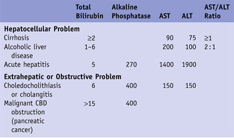
*For clarity, we have chosen typical values that are representative of each condition. Those fields that are empty are either too variable to quantify or not useful as diagnostic guides.


CBD, common bile duct.


IMAGING CONSIDERATIONS


→ Abdominal ultrasound: Least expensive and least invasive; ultrasound should be the first test done for those with jaundice; it visualizes the bile ducts, echotexture of the liver, gallbladder, and pancreas; normal CBD diameter is 4 mm; when ultrasound is performed with Doppler, direction of portal blood flow and presence of thrombi in the portal system can be determined.


$96


→ CT Abdomen and pelvis: Noninvasive; good view of the entire abdominal cavity; requires oral and intravenous contrast for best visualization; patient could have allergy to IV dye or have elevated creatinine that would not allow IV dye to be used, thereby hindering the benefits of the study.


$334


→ MRI/MRCP (MRI/magnetic resonance cholangiopancreatography): Noninvasive; great look at the soft-tissue structures in the abdomen (liver, pancreas, gallbladder); 85% sensitivity for bile duct stones or debris; can’t be done if patients have metal in their bodies or have pacemakers; study can be unfriendly to claustrophobic patients and requires patients to be able to stay still.


$446


→ Endoscopic ultrasound (EUS)–ERCP: Invasive; best method to look at bile ducts. EUS allows for a close-up look at the pancreas, gallbladder, and liver; biopsy via fine-needle aspiration through the EUS scope can be done to sample the liver, pancreas, adrenal gland, and any unusual nodes seen on imaging. ERCP is diagnostic and therapeutic for choledocholithiasis; there is risk of pancreatitis with ERCP.


$885, $2044

Stay updated, free articles. Join our Telegram channel

Oct 3, 2016 | Posted by in MANUAL THERAPIST | Comments Off on Jaundice (Case 25)

Full access? Get Clinical Tree

Get Clinical Tree app for offline access