Fig. 6.1
Shows the intraoperative setup of surgical navigation that includes a stereotactic cameras, a computer station with a monitor showing navigation images and dynamic reference trackers (passive trackers with reflective spheres in this procedure) firmly attached to the patient’s bone and the surgical tool
Navigation involves three essential steps: data acquisition, registration and tracking.
Data Acquisition
Data can be acquired preoperatively and intraoperatively. All medical images are stored in the form of Digital Imaging and Communications in Medicine (DICOM). The preoperative medical images (CT / MR) in the format of DICOM are transferred to the navigation system where image analysis and surgical planning can be performed before the actual operation. The 2D and even 3D fluoroscopic images can be obtained intraoperatively for navigation surgery. Imageless based navigation does not rely on medical images but uses Bone Morphing technique that is a process of recovering the 3D shape of a patient’s anatomy from a few available digitized landmarks and surface points. After surgeons defined specific anatomical bony landmarks using a navigation pointer, a bone model that is derived from the large number of stored CT datasets and fit to the defined bony surface points is generated. These data are then used for registration and tracking.
Registration
Image-to-patient registration is a process in which surgeons tell the computer where the bone is in the space by identifying the anatomical landmarks for the computer. Therefore, it links up the medical images (Xray, CT, MRI or patient’s 3D bone model) with the patient’s anatomy at the operative site. The first method of registration is by placement of tracker pins or fiducial markers at the target bones during 2D/3D image acquisition. Registration is completed after images are acquired or the fiducial markers are identified at the surgery. The second method of registration is surface-matching technique (Fig. 6.2). To start the initial registration, paired-points matching is used to start the initial registration. Four to five predefined points on the preoperative images are matched with the corresponding points on the patient’s anatomy during surgery. To further improve the registration accuracy, more surface points are collected from the target bone that are then matched to the shape of the bone surface model generated from preoperative CT images. The third method is 2D-3D registration (Fig. 6.3). Two fluoroscopic images obtained intraoperatively are matched automatically with preoperative CT images after manual image adjustment.
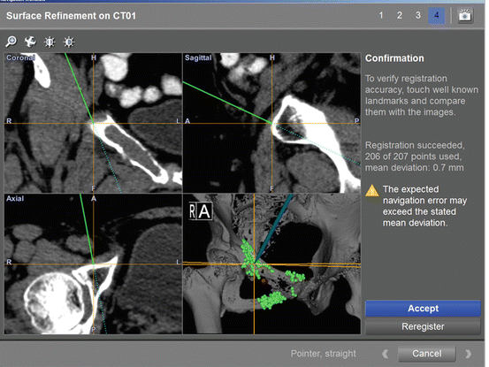
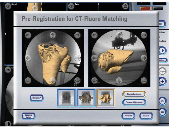
Fig. 6.2
Shows the navigation display after paired-points and surface registration during a CT-based navigated resection in a patient with right pubic malignant tumor. The registration error generated from the navigation machine was 0.7 mm. By placing the navigation probe on the bone surface, the tip of the navigation probe was exactly matching to the preoperative images, thereby verifying the registration accuracy before we can rely on the preoperative images to execute the surgical planning
Fig. 6.3
Shows the navigation display after 2D to 3D registration (CT-fluoro matching) during a CT-based navigated procedure in a patient with proximal tibia tumor. A dynamic reference tracker was first attached to the patient’s tibia. Two fluoroscopic images with different views were then acquired by a navigation-phantom-mounted Xray machine. The two registered images were automatically matched with the 3D bone model generated from the preoperative CT images. This registration method allows registration of preoperative CT images without an open surgical procedure and thus facilitates minimally invasive surgery
Tracking
The target bones and the relative position of surgical tools to the target bone are tracked by 3D optical or magnetic sensors during surgery. Trackers are placed at the target bones while surgical tools are connected with another trackers (Fig. 6.4a, b). The information can be used for identifying the distorted anatomy, visualizing the bone or implant alignment and determining the orientation or level of bone osteotomy.
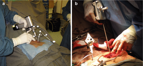
Fig. 6.4
shows passive trackers with reflective spheres (a) and active trackers with infrared light-emitting diodes (b) attaching to the patients’ bones and surgical tools. A stereotactic cameras tracks the target bones and the relative position of surgical tools to the target bones by 3D optical sensors during surgery
Based on the method of referencing information, computer-assisted navigation systems are subdivided into computer tomography (CT) based, fluoroscopic based and imageless. CT-based navigation is the most accurate but it requires additional preoperative CT scanning. It adds extra time for planning and radiation exposure. Fluoroscopic-based navigation is convenient and registered images can be acquired intraoperatively after the placement of patient’s tracker. It is good for trauma fracture fixation and spine surgery in particular with minimally invasive techniques. Imageless-based navigation does not require images. As it does not take into account the unique bony anatomy of each individual, error may arise when surgeons perform the registration by just pointing at bony landmarks.
Clinical Applications and Results
Surgical navigation has been investigated as an adjunct in assisting orthopaedic procedures, particularly in (1) spine surgery; (2) joint arthroplasty; (3) fracture fixation; (4) anterior cruciate ligament reconstruction; (5) bone tumor surgery. In general, use of surgical navigation aims to improve the accuracy of surgical procedures by achieving better position of implants, restoring correct alignment of articular joints or decreasing contaminated tumor resection margin. It is believed that improving surgical accuracy can lead to a better clinical outcome with regards to limbs function, surgical revision rate or tumor recurrence rate. Also, as surgical navigation improves accuracy of surgical instrumentation, it not only may reduce the intraoperative radiation exposure but also allow minimally invasive surgery to be performed.
Spine Surgery
Malpositioning of screws may damage nearby neural and vascular structures in spine surgery, which requires instrumentation. Image guided navigation helps improve the accuracy of screw placement and the surgical safety. It evolves from 2D and then 3D fluoroscopic navigation during surgery. The current intraoperative CT scanner, O arm (Medtronic, Inc., Louisville, CO, USA) allows imaging of the screws intraoperatively.
In a meta analysis of 30 studies with 1973 patients in whom 9310 pedicle screws were inserted, a significantly better accuracy of pedicle screw placement in the cervical, thoracic, and lumbosacral spine was achieved with intraoperative 3D fluoroscopic navigation (95.5 % accuracy) than with 2D fluoroscopic navigation (83.4 % accuracy). Also, both 2D and 3D navigation achieved a better accuracy of pedicle screw insertion than that with traditional fluoroscopy techniques (68.1 % accuracy) [13].
A comparative meta-analysis was carried out by Bourgeois et al. [3]. 3D fluoroscopic-navigated, percutaneous placement of 2132 lumbar pedicle screws were reviewed and compared with 2D fluoroscopic-navigated placement of 4248 screws. The breach rate of screw placement was 0.33 and 13.1 % in 3D and 2D navigated group respectively. 3D fluoroscopic navigation offers markedly improved accuracy of percutaneous lumbar pedicle screw placement in minimally invasive spine surgery.
Joint Replacement Surgery
Surgical navigation was used to increase the accuracy of implant positioning during total knee and total hip arthroplasty as malposition of implant components may compromise implant function and its survival.
Cheng et al. [6] reported a meta-analysis of 41 randomized controlled trials (RCTs) about navigated total knee arthroplasty (TKA). The results suggested that navigation technique improved the accuracy of mechanical leg axis and component orientation. Bauwens et al. [2] did a meta-analysis by reviewing 33 studies (11 RCTs) about navigated TKA with varying methodological quality involving 3423 patients. The alignment of mechanical axes did not differ between navigated and conventional surgery group. However, patients managed with navigated surgery had a lower risk of malalignment at critical thresholds of >3°. Its clinical benefits are unclear as no conclusive inferences could be drawn on functional outcomes or complication rates. Currently, most studies reported that the technique can reduce the outliers of leg alignment and implant malpositioning when compared with conventional technique. There is still no convincing evidence that the marginal benefits can improve the long-term clinical outcome of patient with navigated TKA.
There are few controlled studies examining the role of surgical navigation in total hip arthroplasty (THA). Parratte et al. [18] reported a RCT of 60 individuals undergoing THA with and without imageless computer navigation. There were no significant differences between the two groups in terms of cup anteversion and abduction angles, but with the lowest variations in the navigation group. The authors concluded that the use of imageless navigation can improve cup positioning in THA by reducing the percentage of outliers. Iwana D et al. [11] reported a study of 117 hips undergoing CT-based navigated THA, showing the absolute spatial error of cup position was ≤2 mm for each axis, and the angle error was ≤2° for the cup inclination and anteversion. Current clinical studies suggest that navigation system can help surgeons perform accurate cup placement. CT-based Navigated THA also enabled intraoperative assessment of hip range of motion and limb length [15, 16, 21]. Accurate cup placement was shown to have a better clinical outcome in patients with CT-based navigated THA [22]. In a study with a minimum of 10 years’ follow-up, 46 patients (60 hips) and 97 patients (120 hips) receiving cementless THA with or without CT-based navigation, respectively, were retrospectively reviewed. The navigation group has reduced rates of dislocation and impingement-related mechanical complications leading to revision.
Fracture Fixation
Image-guided navigation has been reported as an adjunct for fixation in pelvic, acetabular fracture as these fractures not only are located at complex anatomical sites but also frequently requires percutaneous screw fixation. The correct entry point and the small target corridor may be difficult to visualize using only an image intensifier.
In a meta analysis of 51 studies including 2353 percutaneous iliosacral screw implantations following pelvic fractures in 1731 patients [27], CT navigation had the lowest rate of screw malposition (0.1 % for 262 screws), but it could not be used for all type of fractures where surgical procedures (reduction maneuvers, additional osteosynthetic procedures) are necessary. The 2D and 3D fluoroscopic navigation and reconstruction techniques provide encouraging results with slightly lower rate of complications 1.3 % (total 445 screws) compared with the conventional technique 2.6 % (total 1832 screws). Rate of screws revision was similar among the three techniques. The study concludes that image-guided navigation is an additional tool to enhance the precision of screw placement and may decrease the rate of revision.
Matityahu A et al. [14] reported a multicenter randomized study of percutaneous sacroiliac screws for pelvic fracture fixation. For 72 patients from the navigated group and 58 patients from the conventional fluoroscopic group, the misplaced screws in navigated and conventional groups are 0 % and 40 % respectively. The authors recommend the use of 3D navigation, where available, for insertion of sacroiliac screws in patients with pelvic fracture.
Ochs BG et al. [17] did a cadaveric study and investigated the role of 3D fluoroscopic navigation in percutaneous periacetabular screw insertion for minimally displaced acetabular fracture. 210 screws were inserted into 30 pelvic models and 30 cadaveric hemipelvis with either conventional 2D fluoroscopy or 3D fluoroscopic navigation. Especially for posterior column screws, due to a lower perforation rate and a higher accuracy in periacetabular screw placement, 3D fluoroscopic navigation procedure appears to be the method of choice for image guidance in acetabular surgery. It remains to be seen whether the favourable results of the navigation technique can be shown in future clinical studies.
Other indication such as intramedullary nailing of long bone fractures has been recently reported by Hawi N et al. [9]. 24 patients who received navigation-assisted treatments and 48 patients who received unassisted treatments, were matched for age, sex, and femur fracture type. Femoral nailing was performed. The results did not support the routine use of navigation in femoral nailing as no improvement in postoperative results or reduction in radiation exposure could be demonstrated when compared with unassisted group. Computer navigation may provide advantages for complicated or sophisticated cases, such as complex 3D deformity corrections.
Related posts:
 Virtual Preoperative Planning
 Computational Image-Guided Technologies in Cranio-Maxillofacial Soft Tissue Planning and Simulation
 Virtual Cranio-Maxillofacial Surgery Planning with Stereo Graphics and Haptics
 Spinal Loading System: A Novel Technique for Assessing Spinal Flexibility in Adolescent Idiopathic Scoliosis
 Navigation in Spinal Surgery
 3D Projection-Based Navigation
Stay updated, free articles. Join our Telegram channel

Full access? Get Clinical Tree


