9 Introduction to MIS Lateral Approach
9.1 Introduction
The lateral minimally invasive surgery (MIS) approach was developed with the goal of reduced morbidity when compared to the anterior and posterior approaches to lumbar interbody fusion.1,2 Compared to the posterior approach, lateral access provides direct visualization of the disk space without requiring direct entry into the spinal canal or retraction of neural elements.3,4 Furthermore, the MIS lateral approach allows for placement of interbody devices with larger footprints.1,5
9.2 Surgical Anatomy
Superficial anatomical landmarks for orientation include the 12th rib, pubic symphysis, and the lateral border of the rectus abdominis. Musculature encountered in this approach, from superficial to deep, includes the external oblique, internal oblique, transversus abdominis, and psoas. Blunt dissection and traversal of the external oblique, internal oblique, and transversus abdominis muscles are not typically associated with significant morbidity, as they are segmentally innervated and denervation is unlikely to occur.
The most significant anatomic concern is transient or permanent neural injury during traversal and retraction of the psoas musculature.6,7 Branches of the lumbar plexus are located within the posterior aspect of the psoas, and consequently these structures can be injured by retraction if they are in close proximity to the surgical corridor (▶ Fig. 9.1). This risk is increased at more caudal levels, with the plexus lying more anteriorly and thus closer to the surgical corridor at the L4–L5 level.8,9 As the risk for neurologic injury is concerning, the use of neuromonitoring during traversal and retraction of the psoas is necessary to identify a safe working zone. Furthermore, the risk of injury to the contralateral retroperitoneal vasculature is also higher at more caudal levels such as L4–L5.8 At these levels, the vasculature is located more posteriorly with greater overlap of the anterior disk space. With greater disk space overlap, the vasculature has a higher likelihood of injury during contralateral annulotomy.
9.3 Surgical Technique
9.3.1 Positioning
Proper positioning is essential during the lateral approach so as to ensure that the surgical plane is perpendicular to the disk space.10 Patients are placed on a radiolucent table in the lateral decubitus position with the operative site positioned over the break in the bed to ensure maximal flexion at the surgical level (▶ Fig. 9.2). In some cases, the table can be retroflexed to increase the distance between the iliac crest and ribs, while the hips can also be moderately flexed to promote psoas muscle relaxation.1,11 The pelvis and thorax are secured with tape and the arms are well padded. Use of an axillary roll also aids in preventing the incidence of brachial plexopathies. The C-arm and surgical monitor should be placed on the side opposite to the surgeon for optimal viewing. Electromyographic (EMG) neuromonitoring is typically used in the lateral approach as branches of the lumbar plexus can be encountered upon traversal of the psoas muscle.10
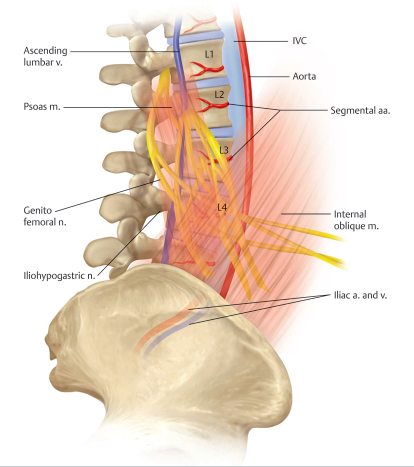
Fig. 9.1 Illustration depicting the positional relationship of lumbar plexus branches and surrounding musculature. (Adapted from An H, Singh K, eds. Synopsis of Spine Surgery. 3rd ed. New York, NY: Thieme; 2016.)
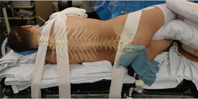
Fig. 9.2 Lateral decubitus positioning for MIS lateral approach. (Adapted from Singh K, ed. Spine Essentials Handbook: A Bulleted Review of Anatomy, Evaluation, Imaging, Tests, and Procedures. New York, NY: Thieme; 2019.)
9.3.2 Approach
The initial descriptions of the lateral lumbar interbody fusion (LLIF) utilized a two-incision approach with a more posterior approach to confirm retroperitoneal positioning prior to deep dissection.1 However, single-incision approaches are now commonly utilized. The lateral incision is placed using fluoroscopic imaging. The abdominal musculature including the external oblique, internal oblique, and transversalis fascia should be bluntly dissected until the retroperitoneum is entered (▶ Fig. 9.3). Once the surgeon has confirmed he or she is in the retroperitoneal space, finger dissection is carried to the lateral surface of the psoas. Serial dilators are then introduced to the disk space through the psoas muscle using fluoroscopy to confirm appropriate placement. Free-running EMG monitoring is used to ensure that the lumbar plexus is not inadvertently injured.12,13 The dilators are then docked to the disk space and secured using a table-mounted retraction system. A fiberoptic light system is then attached to the retractor system to assist with visualization. A self-retaining retractor is then placed, with confirmation of correct localization via anteroposterior (AP) and lateral fluoroscopic views.
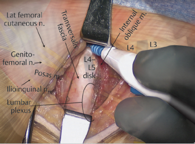
Fig. 9.3 Intraoperative photograph demonstrating dissection of the internal oblique muscle and identification of the transversalis fascia and underlying retroperitoneal space. (Adapted from Singh K, Vaccaro A, eds. Pocket Atlas of Spine Surgery. 2nd ed. New York, NY: Thieme; 2018.)
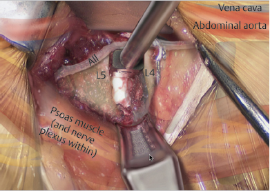
Fig. 9.4 Intraoperative photograph demonstrating endplate preparation. (Adapted from Singh K, Vaccaro A, eds. Pocket Atlas of Spine Surgery. 2nd ed. New York, NY: Thieme; 2018.)
9.3.3 Disk Space Preparation
Bipolar cautery is used to expose the disk space and remove any residual tissue blocking visualization. An annulotomy is then performed, with removal of disk space material using curets, pituitaries, and rasps.14 The annulotomy should be extended to the contralateral side to ensure adequate exposure and correction of coronal plane deformities. The end plates are prepared by removal of cartilaginous tissue using a combination of straight and curved curets (▶ Fig. 9.4).
9.3.4 Interbody Cage Placement
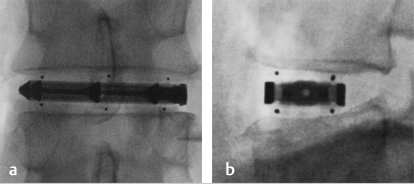
Next, a series of trial sizers are placed under fluoroscopic guidance. Once appropriate sizing has been achieved, the final interbody implant supplemented with an osteobiologic/graft enhancer such as recombinant human bone morphogenetic protein-2 (rhBMP-2) is impacted into the disk space under fluoroscopic guidance. The cage should span the entire width of the vertebral body, thus resting on the ring apophysis and reducing the incidence of subsidence. Final confirmation of cage placement is confirmed via AP and lateral fluoroscopy (▶ Fig. 9.5).