Sterile Instruments/Equipment
- Radiolucent OR table with traction device or fracture table
- 5.0-mm Schanz pins for manipulative joysticks
- Large pointed bone reduction clamps (Weber clamps)
- Ball-spike pushers
- Shoulder hook/bone hook
- K-wires and wire driver/drill
- Reamers
- Implants: depending on the fracture pattern and consistent with the preoperative plan
- Intramedullary nail (trochanteric cephalomedullary nail)
- Screw-side plate device (dynamic hip screw or similar)
- Blade plate (95-degree angle)
- Intramedullary nail (trochanteric cephalomedullary nail)
Patient Positioning
- Supine on a radiolucent table with attachment for sterile traction (see Femur Fracture positioning), or supine on a fracture table.
- Positioning is largely a matter of the surgeon’s preference, with each having advantages and disadvantages.
- For some fractures and in some patients, the use of a fracture table offers predictable indirect reduction and intraoperative imaging and reduces the need for an assistant.
- However, with unstable fracture patterns, the use of a fracture table tends to accentuate deformity in the sagittal plane (usually apex posterior angulation) and posterior translation of the distal fragment(s).
- Some patients have contralateral hip arthrosis and contractures that prevent adequate abduction and flexion to allow quality lateral intraoperative imaging.
- The use of a fracture table has also been implicated in a number of complications to include compartmental syndrome of the contralateral limb, pudendal nerve dysfunction, and perineal skin breakdown.
- Finally, the use of a fracture table requires the presence of a skilled unscrubbed assistant to make intraoperative changes in limb position or traction.
- Prepping the leg free on a flat radiolucent table allows the surgeon unencumbered access to limb position and eliminates complications caused by traction against a perineal post.
- The use of a flat radiolucent table often avoids posterior displacement of the distal fracture fragment as is commonly seen with fracture tables, since the thigh is supported and can be elevated on rolled sterile towels (towel bump).
- However, imaging the hip intraoperatively can be more challenging with intertrochanteric fractures than with other proximal femur fractures.
- For some fractures and in some patients, the use of a fracture table offers predictable indirect reduction and intraoperative imaging and reduces the need for an assistant.
Imaging Tips
- When using the radiolucent flat top table, place a bolster under the ipsilateral sacrum and torso to roll the patient away from the surgeon about 10 to 15 degrees and flex the hip 10 to 15 degrees when the operative limb is placed on a leg ramp.
- This allows for cross-table lateral hip imaging.
- Avoid too large a hip/pelvic bolster so that the cross table lateral of the hip does not demonstrate “relative” neck-shaft retroversion.
- This allows for cross-table lateral hip imaging.
- To adequately image the hip, the C-arm must mirror these positioning modifications.
- The position of the C-arm should be individualized to obtain a true AP image of the hip, with a small crescent of lesser trochanter visible.
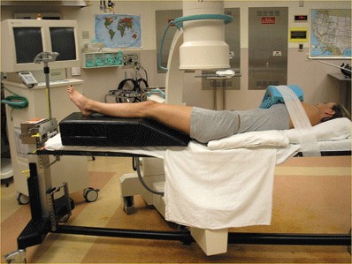
- Often a straight vertical C-arm position with patient bumped up 10 to 15 degrees will offset the anteversion and will give a true AP of the hip (Fig. 14-1).
- The position of the C-arm should be individualized to obtain a true AP image of the hip, with a small crescent of lesser trochanter visible.

Figure 14-1. Patient positioning and fluoroscopic setup for AP view for intertrochanteric fracture treatment.
![]()
- However, some C-arm roll-back may better simulate an AP image.
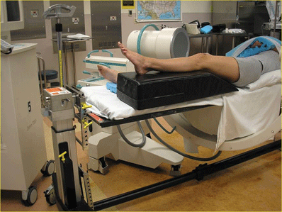
- To obtain a lateral, roll the unit all the way back so that the beam is flat on the floor (Fig. 14-2).

Figure 14-2. Fluoroscope position for obtaining a lateral view.
![]()

Full access? Get Clinical Tree


