Instrumented Lumbar Fusion
Patient Selection
Pedicle screws are anchored in corticocancellous core of vertebral pedicle; strongest point of fixation in spine
Pedicle screws have biomechanical advantage over hooks, wires, and anterior vertebral body screws
Enhance arthrodesis rates in lumbar spine
Important component of transforaminal lumbar interbody fusion, all-posterior correction of idiopathic scoliosis, minimally invasive treatment of fractures, and posterior dynamic stabilization
Indications
Pedicle screws indicated when lumbar fusion performed
Spinal conditions treated using instrumented lumbar fusion include stenosis with spondylolisthesis, spinal tumor, fracture, deformity, iatrogenic instability, and recurrent disk herniation
Contraindications
Absolute contraindications to pedicle screw fixation include pedicle size too small to accept smallest-diameter pedicle screw, congenital absence of pedicle
Relative contraindications include tumor, other bone lesion unable to support screw, profound osteoporosis
Preoperative Imaging

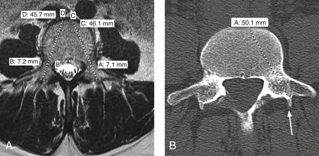
Figure 1Preoperative images used to plan pedicle screw placement. A, Axial MRI shows the templating used to determine the appropriate size of pedicle screws. The widths (lines A and B) and lengths (lines C and D) are determined for pedicle screws bilaterally. B, Axial CT scan shows how to use the accessory process (arrow) as an anatomic landmark for the screw insertion site.
Plain radiographs to help assess spinal alignment; MRI or CT to assess pedicle dimensions.
Note rotational deformities, hyperlordosis, and hypolordosis; all affect screw path
In lumbar spine, visualize starting point for pedicle screw—identified by intersection of line bisecting transverse process horizontally and vertical line along medial aspect of pars interarticularis vertically—on AP view preoperatively
Achieve more precise preoperative planning using axial MRI or CT; assess for factors compromising safe screw placement: dysplastic or absent pedicles, aberrant nerve roots, dural ectasia
Use axial images to measure pedicle diameters, approximate screw lengths at proposed instrumented levels (Figure 1, A); determine screw length preoperatively by measuring from posterior aspect of superior articular process to desired depth within vertebral body (Figure 1, B); use smallest transverse width of pedicle to determine pedicle screw diameter
Do not undersize screws; pullout strength of screw depends on interface between cortical bone of pedicle and screw threads; use screws of slightly larger width as “rescue” screws if screw tract compromised
Careful, slow insertion allows cortical walls to accommodate screws of moderately larger width; no evidence supports using larger-width screws in uncompromised tracts; approximate screw sizes for each level and side transcribed on template paper
Procedure
Room Setup/Patient Positioning
Proper positioning critical; prone position on well-padded radiolucent table; authors prefer Jackson four-poster frame, with chest pad and supports for iliac crest and thighs
Abdomen hangs free to reduce intra-abdominal pressure
Place padding under knees, anterior legs to avoid pressure points
Shoulders abducted, elbows flexed no more than 90° on well-padded arm boards; bolster anterior shoulder to prevent brachial plexus stretch
Head positioner keeps neck in neutral position, avoids pressure around eyes, allows access to endotracheal tube
Secure all wires, lines, catheters to frame of table; allows C-arm to move with less risk of dislodging critical equipment
Table in center of room, anesthesia station at head; surgeon, assistant on opposite sides of patient
Position fluoroscopic imaging system where surgeon can see images; have template paper visible
Before preparing and draping, ensure adequate fluoroscopy can be obtained; adjust table to yield better AP view; use lateral view to mark levels of pedicles to be instrumented to help determine incision size
Special Instruments
Pedicle screws
Stay updated, free articles. Join our Telegram channel

Full access? Get Clinical Tree


