Chapter 7 Injuries about the elbow

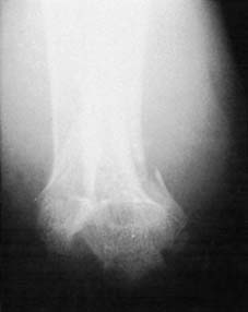
1 Definition: A supracondylar fracture of the humerus is a fracture which occurs in the distal third of the bone. The fracture line lies just proximal to the bone masses of the trochlea (1) and capitellum (capitulum) (2) and often runs through the apices of the coronoid (3) and olecranon fossae. The fracture line is generally transverse.

7 Radiographs (d): With still greater violence there is backward displacement (5), often leading to loss of bony contact. In the AP view there is often medial or lateral shift of the epiphyseal complex (M). The complex may also be rotated relative to the humeral shaft (generally lateral rotation). Rarely, as a result of other mechanisms the distal fragment is displaced and tilted anteriorly (A). Occult fractures may be suggested by the posterior fat pad becoming apparent in the lateral projections.


13 Interpretation of radiographs (d): (6) Reduction is required if there is any significant rotational deformity. This is generally most obvious in the lateral projection. (Left above: no rotational (torsional) deformity present; Right: rotational deformity present.) Rotation, like cubitus valgus and varus, does not remodel well. In this context ‘significant’ is in effect any degree of rotation that is obvious in the plain films.

19 Fixation (a): After reduction, the fracture must be maintained in a stable position. Flexion of the elbow stretches the triceps over the fracture, often splinting it most efficiently. The aim should be to flex the elbow as far as the state of the circulation at the time will permit, while making an allowance in anticipation of further swelling.

25 Check lateral radiographs ctd: This diagram is based on the previous radiograph, and shows correction of all posterior displacement (1), and restoration of the normal angulation of the epiphyseal complex (2). (F = fracture line; C = capitellum; R = radial head.) If a good reduction has been achieved:

27 Absent pulse after manipulation: Returning to Frame 20: If after manipulation the pulse is not palpable, the elbow should be flexed to not more than 100° and maintained in that position with a sling and a back slab (applied over wool and held with a lightly applied bandage). The anaesthetic should be continued while check radiographs are taken.

28 Radiographs (a): If the radiographs show a poor reduction, then no more than two further attempts at manipulation may be permitted under the same anaesthetic. The illustration shows a typical unsatisfactory lateral projection where there is complete loss of contact between the distal fragment and the humeral shaft, with proximal displacement of the distal fragment (compare with Frame 24).

31 Radiographs (d): The preceding case with vascular impairment was remanipulated and the above correction obtained. This is a good reduction, and was accompanied by restoration of the pulse at the wrist.

32 Radiographs (e): A line drawing of the preceding radiograph is shown along with a sketch of the forearm bones drawn in roughly the same position (compare also with Frame 29). (R) = radius, (U) = ulna, (H) = humerus, (F) = fracture, (O) = olecranon, (M) = medial epicondyle.

37 Exploration brachial artery ctd: (iv) The brachial artery (A) is found lying close to the biceps tendon. The median nerve (M) lies on its medial side. The fracture lies deep to the brachialis (B) in the floor of the wound. In some cases relief of local pressure on the vessel (e.g. from the fracture) may restore the circulation, but other measures, requiring experience in vascular surgery, may be necessary.

43 Alternative fixation using a skeletal traction brace: Matuszaki et al2 detail a system which they describe as being easy and safe. A winged screw is inserted into the olecranon, and a spring applies traction. The amount of traction is adjustable by a knurled nut, and the direction of the pull by the position of the traction hook in the wing. Counter traction is provided by a splint which allows for adjustment of the axillary component. The components are radiolucent, making X-ray supervision easy, and it is normally worn for 3–4 weeks.

49 Anterior supracondylar fracture (c): Continuing the traction (1) the elbow is slowly extended (2), reducing the angulation (3). The arm is put in a plaster back slab in a position of 10° flexion (4). This position should not be maintained for more than 3 weeks. At that stage the elbow should be gently flexed (usually about 70–80° can be achieved) and supported either with a sling or further slab until it is united.
Stay updated, free articles. Join our Telegram channel

Full access? Get Clinical Tree


