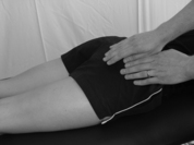
Chapter 1 The hip joint
Anatomy
2. It is an articulation between the head of the femur and the acetabulum of the innominate (hip) bone.
3. It has a strong joint capsule, attaching to the articular margins of the acetabulum and the femoral neck.
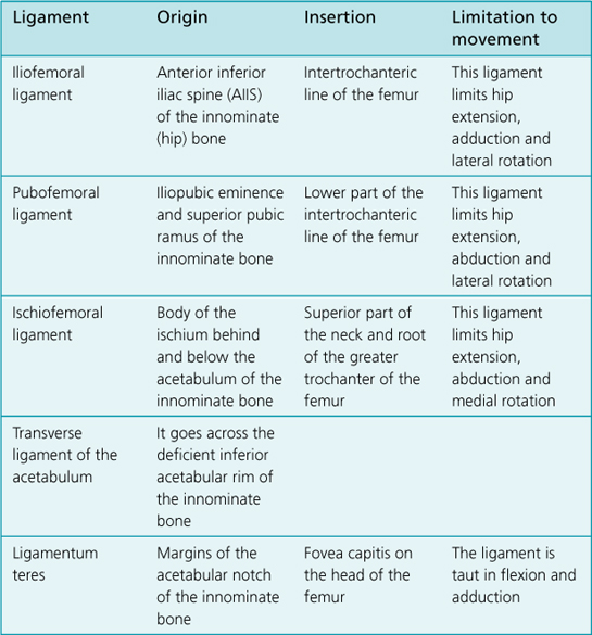
4. It has very strong capsular ligaments – the iliofemoral, pubofemoral and ischiofemoral ligaments.
6. The movements that take place at the hip joint are: flexion, extension, abduction, adduction, lateral (external) rotation and medial (internal) rotation.
Measurement
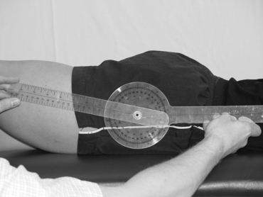
Range of Movement
Extension
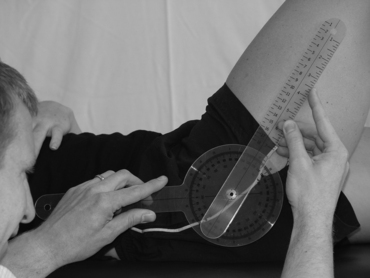
Flexion
Command to patient
‘Bend your knee up towards your chest as far as you can, sliding your heel up the plinth.’
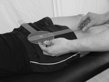
Abduction
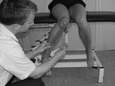
Moveable arm:
This is parallel to the longitudinal axis of the femur, pointing to the middle of the patella.
Command to patient
‘Take your leg out sideways as far as you can. Keep your great toe pointing towards the ceiling.’
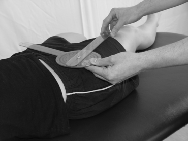
Adduction
Moveable arm
This is parallel to the longitudinal axis of the femur, pointing to the middle of the patella.
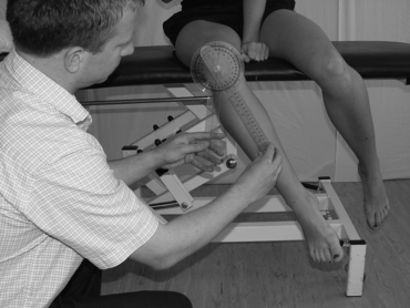
Lateral (external) rotation
Medial (internal) rotation
Muscle Bulk
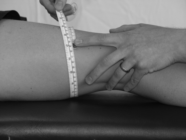
Limb girth: thigh
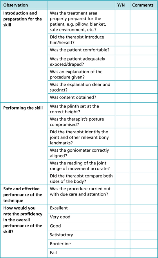
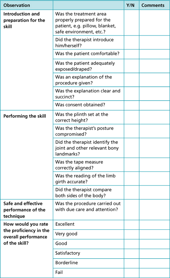
Observational/reflective checklist
Muscle Strength: Oxford Muscle Grading

