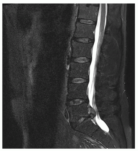
Herniated Disk
Eileen A. Crawford
Nader M. Hebela
CLINICAL PRESENTATION
Intervertebral disks consist of an inner nucleus pulposus surrounded by an outer multilaminar annulus fibrosis. The nucleus pulposus serves to absorb axial loading and transfers this energy to the surrounding annulus fibrosis, a highly organized fiber-reinforced, layered fibrocartilage. The axial load is converted from a compressive force into hoop stresses, allowing energy transfer from the nucleus pulposus and maintenance of disk height, water content, and mechanical function.
Tears in the annulus fibrosis from trauma, degenerative changes, or a combination of both can cause a portion of the gelatinous nucleus pulposus to extrude through the defect of the surrounding annular fibrocartilage. The resultant mechanical pressure exerted on the nerve roots and thecal sac as well as the chemical irritation from direct contact between the nucleus and the nerves lead to signs and symptoms associated with lumbar radiculopathy.
Patients often describe a combination of low back pain and leg pain. They complain of numbness and tingling in the affected lower extremity. The pain may start in the lower back but then travels down the posterior aspect of the buttocks and thighs into the leg and foot. The pain is worse with sitting and is relieved to some degree with standing or lying in certain positions. Patients may experience pain and paresthesias with increasing frequency and duration over days or months. Patients may recall a specific traumatic event that brought on their symptoms, or they may report that the symptoms have gradually progressed over time without a specific inciting event. Patients typically describe the pain as a stabbing pain that starts in their lower back and radiates into the affected buttock, wrapping around the lower extremity in the L4, L5, and S1 nerve root distributions.
Anatomically, the most common sites for a herniated nucleus pulposus occur at the L5-S1, L4-5, and L3-4 intervertebral disks, respectively. The disk herniates posterolaterally in the direction of least resistance, just lateral to the posterior longitudinal ligament. The disk material most commonly causes compression of the traversing nerve roots, although with more superior migration of the herniated disk, the exiting nerve root can also be affected.
While the most common clinical presentation involves one leg or the other, a large enough disk herniation can cause symptoms on both sides of the body. Cauda equina syndrome, an exceedingly rare presentation of disk herniation, may result from a very large compressive lesion that can also affect bowel and bladder function by compressing the entire thecal sac and affecting the S2-4 nerve roots bilaterally.
CLINICAL POINTS
The most common presentation is low back pain with radiating pain, numbness, and tingling into the buttock and down the leg of one side of the body.
The L4-5 and L5-S1 intervertebral disks are the most commonly herniated disks in the lumbar spine.
Patients are usually visibly uncomfortable and prefer standing with the affected knee flexed to decrease the stretch on the sciatic nerve.
PHYSICAL FINDINGS
In the office, patients with a lumbar disk herniation often request to remain standing for the visit, as sitting for even
a short period of time is usually very uncomfortable. They often strive to find a comfortable position to reduce the amount of pain and paresthesias in the affected extremity. They may exhibit decreased lumbar spine range of motion due to pain.
a short period of time is usually very uncomfortable. They often strive to find a comfortable position to reduce the amount of pain and paresthesias in the affected extremity. They may exhibit decreased lumbar spine range of motion due to pain.
Most patients have a near-normal neurologic examination with full motor strength in both lower extremities. Occasionally, patients have weakness in the affected lower extremity muscles innervated by the affected nerve. For example, patients with a right-sided L3-4 disk herniation may not be able to dorsiflex their right ankle with the same strength with which they can dorsiflex their ankle on the left side, indicating compression of the traversing L4 nerve root that innervates the tibialis anterior muscle. While manual muscle testing is useful in determining lower extremity strength, asking patients to simply stand on their toes and then on their heels helps provide the examiner with a better assessment of functional muscle strength. Patients typically experience a difference in sensation between their affected and unaffected sides, reporting that “it just feels like my leg’s falling asleep.” Sensation to light touch is typically preserved. Reflexes on the affected side may be blunted and less brisk than the unaffected contralateral side (Figs. 1-1 and 1-2).
Provocative maneuvers include a positive ipsilateral straight-leg raise test in patients with L4, L5, or S1 nerve root compression. With the patient lying on his back, the examiner flexes the hip to approximately 45 degrees with the knee extended. Dorsiflexion of the ankle places the sciatic nerve on stretch, leading to reproducible pain and paresthesias. A positive contralateral straight-leg raise is considered an even stronger prognostic indicator of a herniated disk. For disk herniations at L2-3, a femoral stretch test can elicit the same pain and numbness that patients normally experience. The patient is asked to lie on the examination table with the unaffected side down. The affected extremity is extended at the hip and flexed at the knee, placing the femoral nerve at maximal tension and eliciting the symptoms the patient has been experiencing.
Stay updated, free articles. Join our Telegram channel

Full access? Get Clinical Tree


