12 Hand
Orientation and general presentation (Figs 12.1 & 12.2)
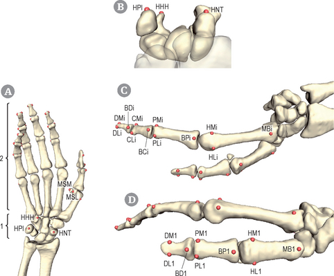
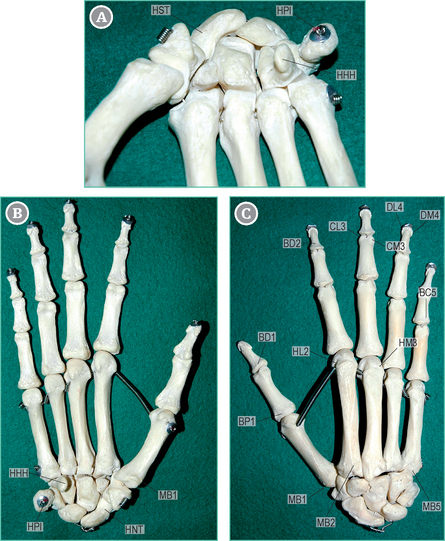
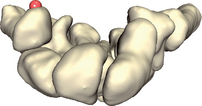
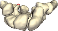
The hand includes carpal bones (1) and digital rays (2), made of long bones (metacarpal bones and phalanges) (Figs 12.1 & 12.2). The most prominent carpal bone is the pisiform (HPI) on the medial aspect of the hand. More distally, the hook of the hamatum (HHH) points forwards. On the other aspect of the wrist, the navicular or scaphoid shows a large tubercle (HNT). Each metacarpus is located on the proximal aspect of a digital ray. A metacarpal bone shows a basis (MBi) and a head, the lateral (HLi) and medial (HMi) sides of which are palpable (i indicates the finger index: 1 = thumb, 2 = forefinger, 3 = middle finger, 4 = annular finger, 5 = auricular finger). Two sesamoid bones, one medial (MSM) and one lateral (MSL), are present on the volar aspect of the metacarpophalangeal joint of the thumb. Distal to each metacarpal bone, three (two for the thumb) phalanges are present in each digital ray. Each phalanx has a basis (BPi, proximal phalanx; BCi, middle phalanx; BDi, distal phalanx) and a head, of which both the lateral side (PLi, proximal phalanx; CLi, middle or central phalanx; DLi, distal phalanx) and the medial side (PMi, proximal phalanx; CMi, middle or central phalanx; DMi, distal phalanx) are palpable. The thumb has no middle phalanx.
Hand/wrist – PIsiform (HPI)[R,L]
Landmark HPI
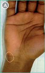
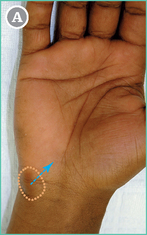
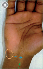
The pisiform is the most prominent carpal bone. It is located on the anteromedial aspect of the wrist (see Figs 12.1 & 12.2).
 The palpator faces the sitting subject, whose elbow is flexed with forearm in supination.
Observe the medial aspect of the subject’s wrist.
The protuberance of the pisiform under the skin is usually visible (orange area).

 Palpate the medial aspect of the wrist until the bony protuberance is found.
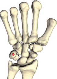
The center of the anterior aspect of the bone is the point to select.
Hand/wrist – Hamatum Hook (HHH)[R,L]
Landmark HHH
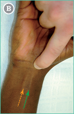
This hamatum is the most medial bone of the second row of the carpal bones (see Figs 12.1 & 12.2). It has a large tubercle (the hook) pointing forwards.
 The palpator faces the sitting subject, whose elbow is flexed with forearm in supination.
First locate HPI (orange area; see p. 92) with your forefinger.
Then glide distally and laterally for about half a pulp-width (blue arrow).
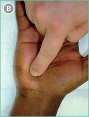
 The hook of the hamatum is found by pressing the pulp into the muscular mass of the hypothenar eminence.
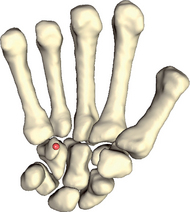
Hand/wrist – Navicular Tubercle (HNT)[R,L]
Landmark HNT
The navicular is the most lateral bone of the first row of the carpal bones (see Figs 12.1 & 12.2). A small tubercle is found on the most distal part of its anterior face.
 The palpator faces the sitting subject, whose elbow is flexed with forearm in supination.
First locate HPI (orange area; see p. 92) with your forefinger.
Stop just lateral to the flexor carpi radialis tendon (running along the green arrow).
Related posts:


Stay updated, free articles. Join our Telegram channel

Full access? Get Clinical Tree


