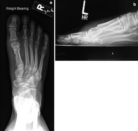
Fig. 4.1
Preoperative anteroposterior (a) and lateral (b) X-ray
4.3 Author’s Preferred Technique
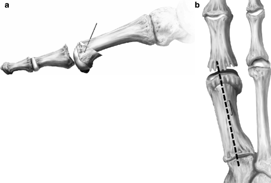
The modified Hohmann procedure is often performed with regional anesthesia and an ankle tourniquet. A dorsomedial incision is performed at the level of the first MP, medial to the long extensor tendon and course proximally more medial to the first metatarsal shaft-neck region. After first MP capsulotomy, dorsal exostoses from both sides of the first MP are removed along with any loose bodies, fractured exostoses, and synovitis. Chondral lesions can be drilled. A pear-shaped burr is used to remodel the first MP. Next, a power saw is used to create an osteotomy at the first metatarsal neck, biased dorsal distal, to proximal plantar, avoiding the sesamoid complex (Fig. 4.2). Often at this point, the capital fragment will “fall” into plantarflexion, reducing the elevatus and excessive length. Usually, the first metatarsal capital fragment is plantarflexed about 5 mm, and shortened to be the same length as the second metatarsal. The osteotomy is temporarily pinned from the proximal first metatarsal into the capital fragment in the desired position. There will be a “dorsal overhang” (that will be removed later).


Fig. 4.2
(a, b) Intraoperative temporary fixation after realignment (plantarflexion) of the distal first metatarsal head (Courtesy Arthrex, Inc., used with permission)
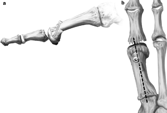
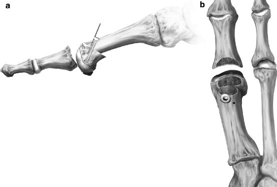
Permanent fixation can be achieved with additional pins or a screw from the proximal first metatarsal shaft into the capital fragment. A counter-sinking pilot hole should be created on the medial surface of the first metatarsal. Often, a 3.5–4.0 mm bioresorbable or metal screw is utilized (Fig. 4.3). The temporary fixation pin can also be replaced with a bioresorbable 1.5–2.0 mm pin to counter any rotation. Any bony overhang is reduced, particularly dorsally (Fig. 4.4). Capsule and skin is closed in standard fashion. Patients are kept non-weight-bearing for 3 weeks in a below-knee cast or cast boot, and weight-bear an additional 2 weeks in the boot. Though it may lag radiographically, X-rays should confirm bony healing (Fig. 4.5). Often patients are clinically healed at 5 weeks post-op. Alternatively, K-wire fixation can be employed; wire removal is performed typically between 4 and 12 weeks when union is achieved (Fig. 4.6). Physical therapy with home exercises begins at 3 weeks. Formal therapy begins at 6 weeks. Return to impact sports is variable but can occur as soon as 8 weeks postsurgery.1


Fig. 4.3
(a, b) Use of bioabsorbable screw to fixate osteotomy (Courtesy Arthrex, Inc., used with permission)

Fig. 4.4
(a) The temporary wire is removed and replaced with a resorbable pin. (b) The dorsal overhang is removed on both sides of the joint. Reduction of dorsal overhang (Courtesy Arthrex, Inc., used with permission)


