Gout
Theodore R. Fields
Making a crystal diagnosis of gout is always ideal, especially because of the possible need for lifelong therapy. Although “minor criteria” for gout exist, crystal identification is always preferred.
Underexcretion of uric acid is the main mechanism of gout, and many medications (such as aspirin and diuretics) worsen this; some medications (such as losartan and fenofibrate) improve the situation.
As the understanding of the natural process of ending a gout attack becomes better understood [e.g., related to transforming growth factor (TGF)-β1 production], improved therapies should follow.
Gout is especially problematic in patients with renal disease—caution is required with nonsteroidal anti-inflammatory agents, colchicine, and allopurinol; each case is individualized regarding decisions to decrease the dose or consider complete avoidance.
The prevalence of gout is increasing and becoming more difficult to treat because of multiple factors, including an aging population, obesity, increased renal disease, and transplantation.
Allopurinol has remarkably improved the life of patients with gout, but there are many patients with allopurinol allergy and renal or hepatic disease who are inappropriate or less-than-ideal candidates for this agent.
Options in patients with allopurinol allergy include oral or intravenous desensitization, uricosuric agents, and the experimental use of oxypurinol, febuxostat, or pegylated uricase.
Patients need clear education about gout and its four stages. Such education will likely improve the recently documented nearly universally poor allopurinol compliance.
Gout is defined as an inflammatory arthritis caused by the deposition of uric acid crystals, which can also be associated with soft tissue urate deposits (tophi) and kidney stones. Gout often accompanies renal disease, hypertension, and coronary artery disease, but the causative role of uric acid in these conditions remains under active investigation. Modern treatment has remarkably reduced the suffering and complications of gout, but there
are still many patients who need new agents for the management of this very painful and disabling disorder.
are still many patients who need new agents for the management of this very painful and disabling disorder.
Primary gout (Table 43-1) is due to genetically determined hyperuricemia, which is caused by urate overproduction or underexcretion. Enzyme defects causing urate overproduction, in a small percentage of patients with gout, have been well worked out. Underexcretion, found in most cases, has recently been tracked to hereditary defects in renal anion transporters of uric acid.
Secondary gout (see Table 43-1) can be due to intake of medications that decrease urate excretion or cause renal insufficiency, increased purine intake in the diet, and treatments or conditions that lead to cell breakdown and consequent purine production. Uric acid is the end product of purine degradation.
Uric acid crystals induce a classical inflammatory response via an activation of the innate immune system. Toll-like receptors are involved in recognizing urate crystals; complement, including the membrane attack complex, is activated; neutrophils and then macrophages are recruited into the joint via chemotactic factors; and cytokines [e.g., tumor necrosis factor-α (TNF-α), interleukin (IL)-1, and IL-8] are secreted.
Gouty attacks are self-limited because of the nature of the immune system response to urate. This is related to the fact that neutrophils eventually stop entering the joint, and as monocytes mature within the joint fluid, they have been found to produce fewer proinflammatory cytokines.
URIC ACID, THE HEART, AND KIDNEY. It is clear that hyperuricemia correlates with renal disease and atherosclerosis, but it remains unclear as to what extent, if any, hyperuricemia is directly causative of each. Urate level is routinely elevated in the metabolic syndrome (i.e., insulin resistance, hypertension, central obesity, dyslipidemia, and the proinflammatory and prothrombotic state), at least in part because of high insulin levels, which decrease urate excretion. It is presently unclear whether lowering urate levels in the metabolic syndrome will ameliorate any of the clinical features of this syndrome.
Table 43-1 Causes of Hyperuricemia
Overproduction (10%)a
Underexcretion (90%)
Primary overproduction:
Primary underexcretion:
- Hereditary enzyme defects:
–HGPRT deficiency
–PRPP synthetase overactivity
Secondary causes of overproduction:
- Ethanolb
- Myeloproliferative disorders
- Cytotoxic chemotherapy
- Sickle cell anemia
a Underexcretion is essentially universal in gout—present even in overproducers—but overproducers are the patients with hyperuricosuria and worth identifying.
b Ethanol both increases urate production and decreases its excretion.
c This increase in urate has recently been reported to be transient and of small magnitude.
HGPRT, hypoxanthine-guanine-phosphoribosyltransferase; PRPP, phosphoribosyl pyrophosphate; URAT1, urate transporter.- Hereditary enzyme defects:
The prevalence of gout is increasing, and there is a trend toward an earlier age of onset. Gout is the most common inflammatory joint disease in men older than 40 years.
Men continue to outnumber women in the incidence of gout, but after menopause, women begin to close the gap, possibly related to the loss of the uricosuric effect of estrogen.
Attacks of gout are becoming more severe and difficult to treat. This likely relates to the aging of the population, with their comorbidities contraindicating many of our therapeutic options. Aging patients have a higher incidence of renal disease and renal failure, which worsens hyperuricemia and limits the use of many of our standard agents. In patients undergoing transplantation, cyclosporine can cause an especially aggressive form of gout, with rapidly forming tophi.
Four different stages are recognized in the clinical evolution of gout.
Asymptomatic hyperuricemia. Although no attacks of gout occur in this phase, urate deposits can develop in the joints, and in some patients, their 24-hour urinary urate excretion rate is high enough to increase the risk of kidney stones.
Acute gouty attacks. In this stage, the patient develops painful, inflamed joints and almost always seeks treatment.
Intercritical gout involves periods between attacks in a patient who has already had at least one attack.
Advanced and tophaceous gout is a stage in which patients develop chronic abnormalities, such as chronic arthritis with superimposed flares, x-ray changes in joints, and tophi (visible nodular collections of uric acid) (Fig. 43-1).
Attacks of gout are generally of very rapid onset, often with systemic features, such as low-grade fever. After a first attack, the second may not occur for 5 to 10 years in some patients, but once the attacks begin to occur frequently, a pattern of closely spaced attacks is almost always established, and long-term spontaneous remission is rare. The classic attack of gout involves only a single joint, but as the disease evolves, it is not rare to see polyarticular attacks and involvement of contiguous joints.
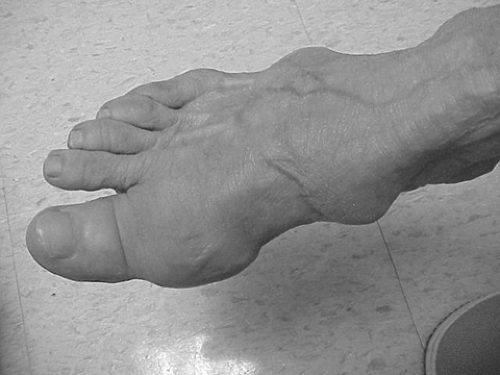
Figure 43-1. Gouty tophi.
Table 43-2 Making the Diagnosis of Gout
One major criterion is sufficient:
- Urate crystals in a joint or a proven tophus
Presence of six of 12 minor criteria:
- More than one attack of acute arthritis
- Maximal inflammation developed within 1 d
- Attack of monarticular arthritis
- Joint redness observed
- First metatarsophalangeal joint painful or swollen
- Unilateral attack involving first metatarsophalangeal joint
- Unilateral attack involving a tarsal joint
- Suspected tophus
- Hyperuricemia
- Asymmetric swelling within a joint (radiograph)
- Subcortical cysts without erosions (radiograph)
- Negative culture result of joint fluid for microorganisms during an attack of joint inflammation
Adapted from Wallace SL, Robinson H, Masi AT, et al. Preliminary criteria for the classification of acute arthritis of primary gout. Arthritis Rheum 1977:20;895–900, with permission. Related posts:

Stay updated, free articles. Join our Telegram channel

Full access? Get Clinical Tree


