Sterile Instruments/Equipment
- Sterile drapes, including impervious stockinette and 4 inch elastic bandage wrap for forearm and hand
- 1/4% bupivicaine, with epinephrine for posterior incision (minimizes skin bleeding)
- Large and small pointed bone reduction clamps (“Weber clamps”)
- Implants
- 2.7- and 3.5-mm reconstruction plates and screws
- 1/4 and 1/3 tubular plates
- 2.0- and 2.4-mm plates and screws
- Extra long screws (2.0, 2.4, 2.7, and 3.5 mm)
- 2.7- and 3.5-mm reconstruction plates and screws
- K-wires and wire driver/drill
- Femoral distractor(s) if reducing displaced fractures, especially if >20 days after injury
Positioning
- Lateral: modified Judet approach
- Lateral position is preferred to prone.
- The lateral position allows for better palpation of the coracoid for screw placement, percutaneous “joystick” placement in the coracoid as needed, and facilitates reduction maneuvers.
- Place the patient on the cantilevered end of a radiolucent operating table (reversed) which facilitates intraoperative imaging.
- Chest roll for axillary protection, using a beanbag and appropriate padding.
- A padded Plexiglas board supports the uninjured, dependent arm; the operative arm is prepped free and supported on a padded (pillow) sterile Mayo stand.
- This enables intraoperative limb manipulation and positioning to aid in reduction and imaging.
- Pad Mayo stand and tray with a taped pillow; cover with a sterile Mayo stand cover.
- This enables intraoperative limb manipulation and positioning to aid in reduction and imaging.
- Alternatively, the operated upper extremity may be supported by an adjustable radiolucent arm board under the drapes (Figs. 2-1 and 2-2).
- Fluoroscopy should be positioned perpendicular to the patient and table, entering from the patient’s anterior/front side, opposite from the surgeon.
- This allows a rollover image for a scapular Y view, and a rollback to image a Grashey AP view.
- Lateral position is preferred to prone.
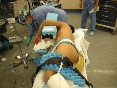
Figure 2-1. The lateral decubitus position for Modified Judet surgical approach.
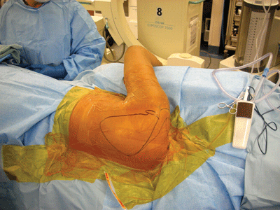
Figure 2-2. Prepped and draped. The arm is prepped into the field so it can be manipulated sterilely. There should be a clear path for the C-arm to come in from the opposite side.
Surgical Approach
- Modified Judet.1
- The incision is curvilinear, and parallels the scapular spine and the medial scapular border (Fig. 2-3).
- Prior to incision, injection of Bupivicaine 0.5% with epinephrine can help minimize cutaneous bleeding.
- Develop a full thickness skin and subcutaneous flap, taking care to leave the fascia intact over the posterior scapular muscles and latissumus dorsi (Fig. 2-4).
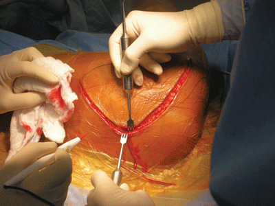
Figure 2-3. Curvilinear incision.
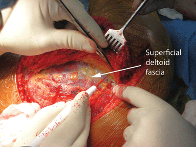
Figure 2-4. A full thickness flap is elevated from the deltoid fascia.
![]()
- Identify the lower border of the deltoid, and incise the fascia sharply (Fig. 2-5A,B).
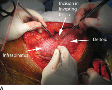
Figure 2-5. A,B: The investing fascia is incised along the inferior border of the posterior deltoid.
![]()
- Separate the inferior border of the posterior deltoid from the infraspinatus from medial to lateral.
- Keep the deep fascia with the deltoid as reflected (Fig. 2-6).
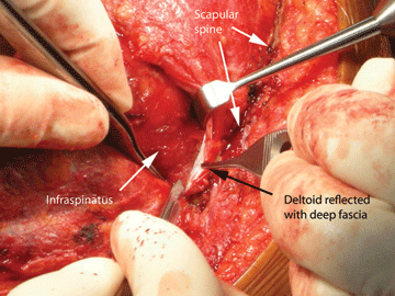
Figure 2-6. The deltoid is reflected from the infraspinatus with the deep fascia.
![]()
- Sharply release the deltoid origin from the scapular spine.
- A tag stitch in the superomedial corner aids in retraction and identifies this area for later repair (Fig. 2-7).
Related posts:


Stay updated, free articles. Join our Telegram channel

Full access? Get Clinical Tree


