1 General organization of the cardiovascular system
1.1 Introduction to the cardiovascular system
The cardiovascular system has many jobs:
• The cellular distribution of oxygen and nutrients (amino acids, fatty acids, vitamins)
• The elimination of cellular waste (carbon dioxide, lactates)
• The transport of oxygen, carbon dioxide, and hormones
• The regulation of body temperature, blood pH, water volume, mineral salt levels, etc.
1.1.2 Vascular network
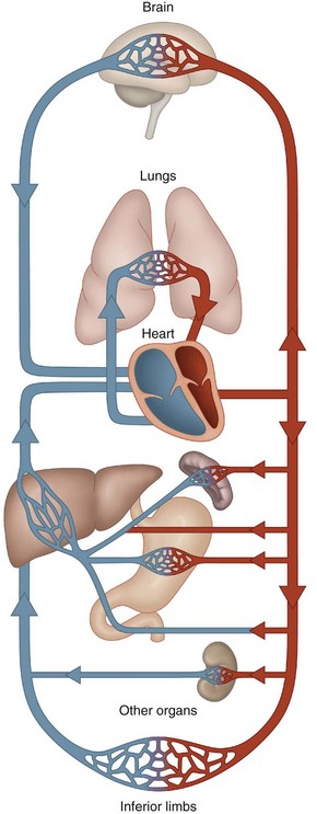
When looked at from an anatomical rather than a functional point of view, the vascular circuit can be divided in two (Fig. 1.1):
• The systemic or great circulation carries oxygen and nutrients to the tissues. The systemic circuitry feeds the organs of the body through a series of blood vessels arising from the left ventricle via the aorta. The systemic veins collect blood from these organs and convey it back to the right atrium of the heart.
• The pulmonary or small circulation denotes the shuttle between the heart, which generates the flow, and the lungs, which provides the oxygen.
1.1.3 The heart
1.1.4 Vascular sections – resistance and capacitance
High pressure system
Several subcompartments can be distinguished:
• The reservoir vessels comprise the aorta and its large branches. Their walls are elastic and they convert, without notable modification in average pressure, the intermittent pressure of the cardiac pulsation into a smooth type of flow. These are the main vessels of blood distribution to the periphery.
• The arterioles are the site of regional resistance and are the regulatory vessels of blood flow.
• The precapillary sphincters are smooth muscle bands that lie ‘before’ the capillaries and determine the flow at the entrance to the capillaries in order to control perfusion.
1.1.5 Blood distribution
At rest
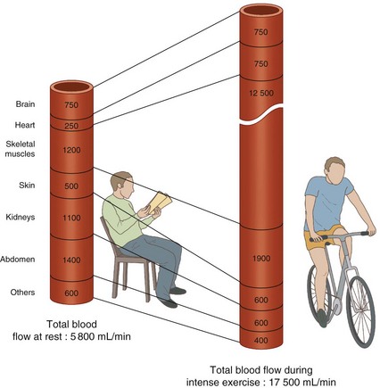
At rest, the digestive system and the kidneys attract about 50% of the blood distributed, whereas the heart brings in only 3–4% (Table 1.1 & Fig. 1.3).
| Organs | % total |
|---|---|
| Digestive | 25 |
| Kidneys | 20 |
| Brain | 13 |
| Skin | 9 |
| Heart | 4 |
With exertion
During heavy muscle work, the cardiac output increases to 17–25 L/min. Distribution to various tissues is modified, with the exception of the brain, whose supply remains remarkably constant (Table 1.2 & Fig. 1.3).
Table 1.2 Blood flow on exertion
| Organs | % total |
|---|---|
| Heart | 4 |
| Brain | 14 |
| Kidney | 20 |
| Skeletal muscle | 22 |
| Skin | 8 |
| Other organs | 12 |
1.1.6 Types of circulation
Depending on the purpose of the blood running through an organ, three types of circulation occur:
• Nourishing circulation: cerebral arteries, muscular arteries, coronary arteries, bronchial arteries, hepatic arteries
• Functional circulation: pulmonary arteries, portal vein
• Mixed circulation: renal arteries, mesenteric arteries, cutaneous arteries.
1.2 The heart
1.2.1 Anatomy review
Form and orientation
On the outside, the heart is a somewhat rounded conical form, lying on its side.
• The apex of the heart is directed caudad, anteriorly and to the left.
• The base of the heart, opposite the apex, is directed cephalad, and faces mainly posteriorly and to the right.
• The main axis of the heart is oblique from posterior to anterior, from right to left. This obliqueness varies with the shape of the thorax; the heart lying more vertically in a narrower thorax.
Chambers
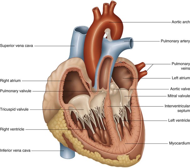
The hollow muscle of the heart is divided into right and left parts by the interatrial septum. Each side of the heart has two chambers, an atrium (superior chamber) and a ventricle (lower chamber) connected by atrioventricular valves (Fig. 1.4).
Atria
Three veins enter the right atrium:
Four pulmonary veins, two right and two left, enter the left atrium.
Structure of the heart
Myocardium
• striated muscle fibers anchored to a fibrous framework
• differentiated fibers forming the nodal tissue or conducting system of the heart.
Muscular fibers
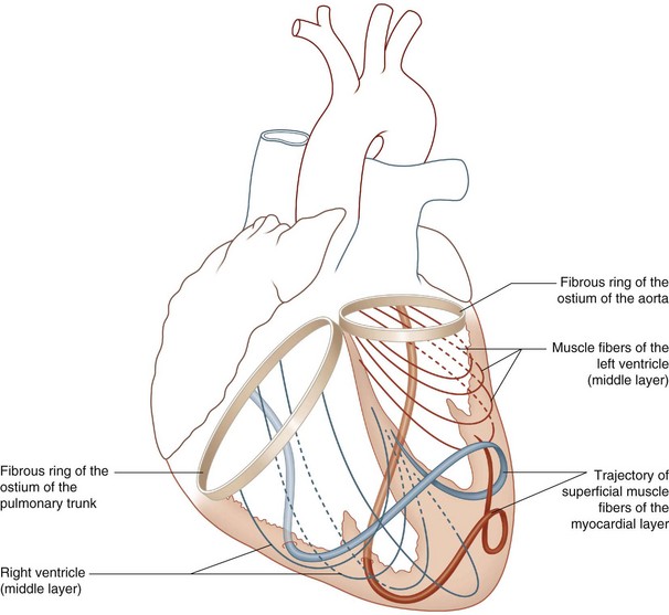
The heart is composed of ventricular and auricular fibers (Fig. 1.5).
The ventricular fibers consist of:
• fibers specific to each ventricle whose oblique moorings attach to the fibrous rings of the skeleton
• Common fibers that envelop and join the two sacs formed by the individual fibers.
Conduction system of the heart
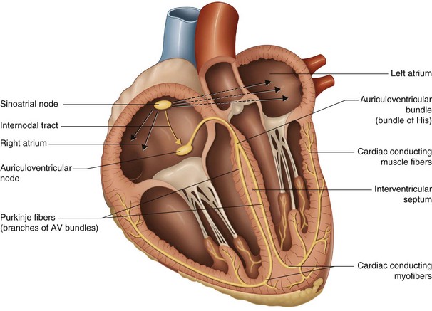
The heart has an intrinsic system by which it is able to beat without the participation of external nerves. This property is termed autorhythmicity. This autonomous command system is the tissue nodal or impulse conducting system (Fig. 1.6) made up of three parts:
• The sinoatrial (SA) node (Keith and Flack) is located in the wall of the right atrium, at the junction of the superior vena cava and the external atrial orifice. This is the pacemaker of the heart.
• The atrioventricular (AV) node (Aschoff–Tawara) is a smaller collection of nodal tissue situated in the interauricular septum, near the atrioventricular valves. It functions as a secondary pacemaker.
• The atrioventricular (AV) bundle (of His) is a collection of specialized muscle fibers beginning at the AV node and dividing into right and left bundles at the junction of the membranous and muscular parts of the interventricular septum.
• The AV bundles proceed on either side of the septum, deep to the endocardium, and then ramify into the subendocardial branches (Purkinje network), which extend into the walls of the respective ventricles.
Pericardium
• The serosal pericardium: this glistening serous membrane facilitates all manner of movements, glidings, and deformations of the heart, in relation to its neighboring organs.
• The fibrous pericardium clothes the serous pericardium. The heart is protected and somewhat tethered in place inside this fibrous sac.
Stay updated, free articles. Join our Telegram channel

Full access? Get Clinical Tree


