Galeazzi Fracture-dislocation
25.1 Introduction
Fracture of the shaft of the radius complicated by dislocation or subluxation of the distal radioulnar joint (DRUJ) is commonly referred to as Galeazzi fracture-dislocation. DRUJ involvement is the unique feature of this type of injury, which accounts for nearly 7% of all fractures of the forearm in adults and nearly 3% in children. However, the true incidence remains unknown because Galeazzi lesions are frequently underdiagnosed. Misdiagnosis or inappropriate treatment will result in persistent DRUJ instability and wrist pain, as well as decreased grip strength and forearm rotation.
Asley Cooper was the first to describe a distal radial shaft fracture with disruption of the DRUJ in 1824.1 However, this injury has become connected with the name of Ricardo Galeazzi who in 1934 reported his experience with 18 such cases.2 It is also known as reverse Monteggia fracture, Piedmond fracture, or Darrach–Hughston–Milch fracture, while the term “fracture of necessity” is also frequently used to describe this inherently unstable injury that requires surgical treatment to achieve favorable outcomes, particularly in adults. The term Galeazzi-equivalent lesion was introduced in 1982 to describe a fracture of the distal radius in association with a fracture of the distal pole of the ulna in adults or separation of distal ulnar epiphysis without DRUJ disruption in children.3
25.2 Mechanism of Injury
Galeazzi fracture-dislocation may occur during a fall and less frequently during motor vehicle accidents, electric shock, or blunt trauma. It is the result of forceful axial loading of the maximally pronated forearm while the wrist is in extension. The deforming forces include those of the brachioradialis, pronator quadratus, and thumb extensors. These deforming muscular forces cannot be controlled by conservative treatment with plaster immobilization. According to some authors, axial loading of the maximally supinated forearm can also result in the same type of injury.4,5
25.3 Pathophysiology
Important structures in the development of Galeazzi fracture-dislocations are the DRUJ, the radius, and the interosseous membrane that runs in oblique fashion from the radius to the ulna and firmly constrains these two forearm bones. The direction of the interosseous membrane fibers helps to prevent radial shortening. However, the absence of any attachment of the interosseous membrane in the distal one-third of the radius allows shortening of the latter when fractures occur in this part of the radius. Moreover, the junction of the middle and distal one-thirds of the radius is at increased risk of fracture due to unique bone mineral content and cross-sectional geometrical properties of the radius at that level.6
In a cadaver study, Moore et al7 showed that the fracture of the radius is preceded by the lesion of the interosseous membrane and triangular fibrocartilage complex (TFCC). The same authors demonstrated that shortening of the radius by less than 5 mm did not result in DRUJ disruption, whereas shortening of more than 10 mm was associated with both TFCC and interosseous membrane tears. Mikić8 supported the idea that rupture of the TFCC is the main cause of the DRUJ redislocation.
Instability of the DRUJ may occur due to failure to recognize the injury, failure to reduce the dislocation intraoperatively, nonanatomical radial reduction, or interposed soft tissue that blocks reduction. Isolated fractures at the distal one-third of the radius are not always associated with DRUJ dislocation, although the fractured radius shortens and tends to cause subluxation of the DRUJ and dorsal angulation of the radius. On the other hand, DRUJ disruption is often identified in patients with fractures of the middle and proximal thirds of the radius diaphysis.
Predictors of TFCC injury and consequent DRUJ instability following a distal radius fracture include the following:
The magnitude of fracture displacement
The fracture pattern
The magnitude of DRUJ widening
The presence of an ulnar styloid fracture
According to Fujitani et al9 the strongest independent factor for instability is the DRUJ widening. An increase in the DRUJ distance by 1 mm is associated with a 5-fold increase in the risk of tear in the radioulnar ligament, a primary stabilizer of the DRUJ.
25.4 Imaging
A radiographic examination of the DRUJ is crucial for the diagnosis of Galeazzi fracture-dislocation. Standard anteroposterior and true lateral forearm views should be obtained. Radiographic evaluation must also include anteroposterior and lateral views of the wrist, along with anteroposterior and lateral views of the elbow. Radiographs of the uninjured contralateral wrist are also required for comparison. The surgeon must always be alert for specific findings on radiographs (▶ Fig. 25.1a, b), such as
widening of the DRUJ space
fracture at the ulnar styloid base
subluxation or dislocation of the ulna relative to the radius on a lateral radiograph
shortening of the radius by more than 5 mm relative to the distal ulna
asymmetry compared with the contralateral DRUJ.
Computed tomography is indicated for the diagnosis of DRUJ disruption when plain films are inconclusive. Magnetic resonance imaging evaluation and/or wrist arthroscopy may reveal DRUJ disruption or TFCC tear, although it is not routinely undertaken.
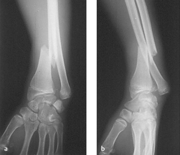
Fig. 25.1 (a) Anteroposterior and (b) lateral views of a Galeazzi fracture-dislocation reveal dislocation of distal radioulnar joint, and a short oblique fracture at the distal one-third of the radius with more than 5 mm of shortening.
25.5 Classification
Different classification systems have been proposed by several authors for description of Galeazzi fracture-dislocation. Mikić7,8 was the first to describe five types of radial shaft fracture based on the location of the fracture along the axis of the radius: (a) at the junction of the middle and distal one-thirds of the radius; (b) in the middle one-third of the radial shaft; (c) at the junction of the proximal and middle one-thirds; (d) the distal one-third; and (e) the proximal one-third of the radius. However, his study did not focus on the presence of DRUJ instability in relation to the location of the fracture.
Based on the anatomical location of the fracture in the radius shaft, Maculé Beneyto et al10
Related posts:
 Undisplaced Scaphoid Fractures
 Proximal Row Fractures (Other Than Scaphoid and Pisiform Fractures): Triquetrum and Lunate Fractures
 Intra-articular Fractures of the Distal Radius (AO types C3, with Special Focus in C3.3), Open Approach
 Fracture-dislocations Other than Perilunate
 Intra-articular Fractures of the Distal Radius (AO type C3), (Dry) Arthroscopic Approach
 Unstable Ulnar Styloid Fracture
Stay updated, free articles. Join our Telegram channel

Full access? Get Clinical Tree


