Fig. 1.1
The radial head forms an angle of 165° with the radial neck axis
Thanks to this conformation, the radial head is positioned in the frontal plane.
The superior concavity, ovoid shape and head size vary from one individual to the other.
It has a sponge-like structure so it’s fragile but reinforced by bone trabeculae whose homogeneous distribution avoids the formation of areas more fragile than others.
In supination, the ovoid axis is oriented inwards and frontwards, and the radial tuberosity is oriented inwards. The radial head tilts so that its internal part is higher than its external part.
Lesser Sigmoid Cavity (or Radial Notch)
It’s a small cavity with a cartilaginous “crust” located under the greater sigmoid cavity (or trochlear notch). Both notches are separated by an anteroposterior ridge.
1.1.1.2 Diaphysis
The radial and ulnar diaphyses have special curvatures that allow important mobility in pronosupination.
In supination, the two bones are concave anteriorly but have opposite concavities in the frontal plane, where the ulna is concave outwards and the radius is concave inwards (Fig. 1.2).
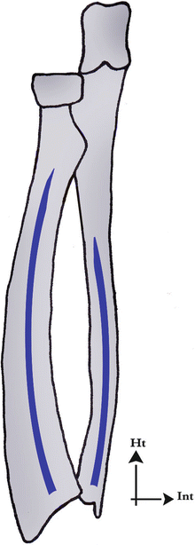
Fig. 1.2
The diaphysis of the two bones of the forearm have inverted concavities in the frontal plane, which allows their “winding” in pronation
1.1.1.3 Distal Extremities
Along with the triangular complex, they form a set that interfaces with the 1st carpal row.
Radial Epiphysis
It has the shape of a truncated pyramid whose anterior part is occupied by the pronator quadratus. Its posterior side contains Lister’s tubercle, which separates two grooves: the extensor pollicis longus passes and changes direction in the medial one and the radial extensors of the carpus pass in the lateral one. On its lateral side, there is the radial styloid process, in front of which pass the tendons of the abductor pollicis longus and extensor pollicis brevis.
The inferior articular side has a medial part (relation with the lunate) and an external part (relation with the scaphoid). It is concave and globally oriented frontwards (10°) and inwards (25°) (Fig. 1.3).
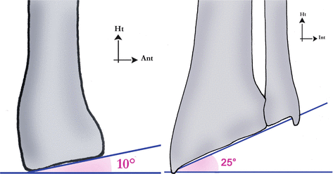
Fig. 1.3
Orientation of the radial socket downwards, frontwards and inwards
The internal side receives the radial notch of the ulna (trochoid joint).
Mechanically, the radius absorbs 80 % of the axial constraints transmitted by the carpus. A 2 mm shortening is enough to transfer 20 % more load on the ulnar compartment.
Ulnar Epiphysis
Thinner than the radial epiphysis, it contacts with the radius by its external convex side to form the distal radioulnar joint.
Its inferior side has to do with the triangular complex, which articulates with the 1st carpal row.
1.1.2 1st Carpal Row (Fig. 1.4)
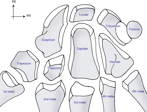
Fig. 1.4
Carpal bones
The mobility between the bones of the 1st carpal row is complex and gives it a huge adaptive capacity allowing it to absorb part of the constraints imposed on it. From lateral to medial, it’s made of:
1.1.2.1 Scaphoid
The scaphoid has a “bean” shape. It’s the most lateral bone of the 1st carpal row. There are an angle of 30° between the distal and proximal parts viewed from the side and an angle of 40° viewed from the front (Figs. 1.5 and 1.5′).
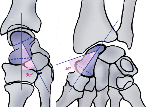
Figs. 1.5 and 1.5′
Intra-scaphoidal angles
More than 75 % of its surface is covered by cartilage.
It’s the most voluminous bone of the 1st carpal row and the closest to the thumb, between the radius and the trapezo-trapezoidal joint.
Like for the lunate and triquetrum, there are no tendons attached on it, but it’s strongly attached to the radius and carpus by intrinsic and extrinsic ligaments.
It forms a 45° angle with the radial axis in the frontal and sagittal plane. This orientation is the key element of the natural thumb opposition, essential in prehensions (Figs. 1.6 and 1.6′).
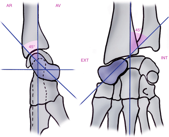
Figs. 1.6 and 1.6′
Orientation of the scaphoid frontwards and outwards with respect to the radial axis
It’s made of three parts (Fig. 1.7):
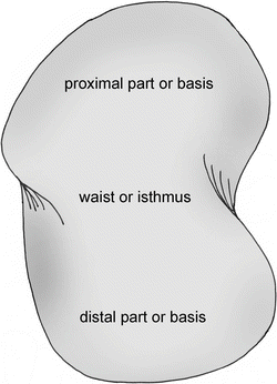
Fig. 1.7
The three parts of the scaphoid
The proximal part is round, covered in cartilage and articulates with the lunate (plane joint). This joint forms the lateral part of the carpal condyle. The superior side of the proximal part articulates with the radius. It can be palpated in wrist flexion, when it dorsally peeks out from the radiocarpal groove in the Lister’s tubercle axis.
The neck is the narrowest part of the bone (6 mm width). Its medial part articulates with the capitate. Its lateral part contacts with the radial artery and corresponds to the floor of the anatomical snuffbox.
The base articulates with the trapezium and trapezoid distally and is anteriorly prolonged by the scaphoid’s tubercle. On this tubercle are inserted the flexors retinaculum and the abductor pollicis brevis. There’s a medial groove where the flexor carpi radialis passes. This muscle then ends on the scapho-trapezo-trapezoidal joint and on the bases of the 2nd and 3rd metacarpals. It’s easily palpable in wrist extension.
Its vascularization is ensured by the branches of the radial artery, in two groups:
The proximal group enters the scaphoid’s neck and takes care of 70 % of the vascularization.
The distal group enters the scaphoid’s tubercle and takes care of 30 % of the vascularization (Fig. 1.8).
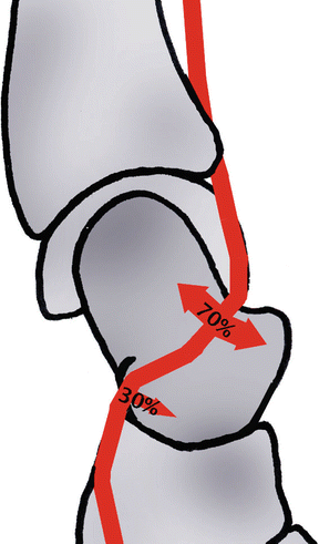
Fig. 1.8
The vascularization of the scaphoid is ensured by two groups given off by the radial artery. The proximal group enters by the waist and represents 70 % of this vascularization. There are no vessels that directly enter the head of the scaphoid
There are no vessel entering the proximal part, which explains why there is an important risk of pseudoarthrosis and necrosis in case of fractures at this level.
The scaphoid is “stuck” between the radius and the trapezo-trapezoidal joint and is under important compressive constraints as 80 % of the carpus load is transmitted to the radius and 20 % to the ulna. The scaphoid transmits 60 % of these constraints and the lunate 40 % (Fig. 1.9).
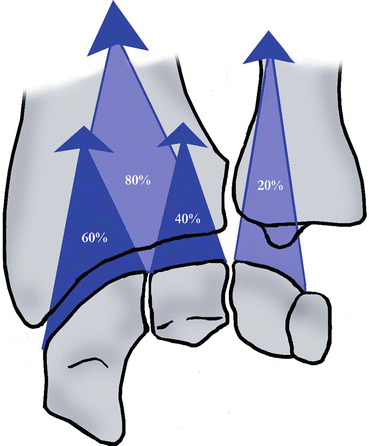
Fig. 1.9
The radius bears 80 % of the longitudinal forces coming from the carpus. The scaphoid transmits 60 % of these forces to the radius
These axial constraints tend to bring the scaphoid to flexion because of its angle of 45° with the radius (Fig. 1.10).
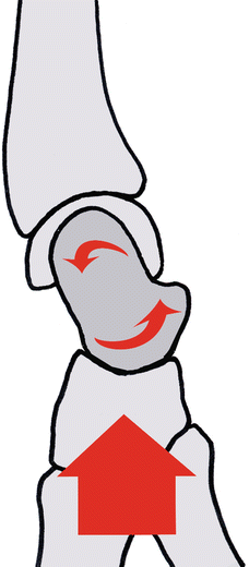
Fig. 1.10
The scaphoid’s orientation with respect to the radius in the sagittal plane causes a flexion of the radius because of the axial constraints coming from the carpus
However, several anatomical elements oppose this tendency (Fig. 1.11):
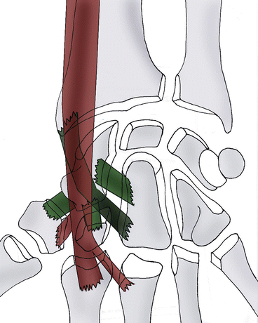
Fig. 1.11
Ligamentary (scapho-trapezo-trapezoidal system, scaphocapitate ligament, radioscaphocapitate ligament) and muscular (flexor carpi radialis) elements opposing the flexion of the scaphoid
The scapho-trapezo-trapezoidal ligamentous system and the scaphocapitate ligament (powerful distal anchor)
The radioscaphocapitate ligament that opposes the distal part of the scaphoid from going forward
The flexor carpi radialis, sliding in front of the scaphoid and whose contraction brings the distal part of the scaphoid backwards
1.1.2.2 Lunate
It has the shape of an irregular lunar crescent with a distal concavity articulated with the capitate. Its anterior horn is bigger than the posterior one. Its lateral side articulates with the scaphoid and its internal side with the triquetrum.
When the wrist is straight, the two horns are at the same height, and the lunate axis is the same as the radial axis.
It’s subjected to two opposite forces through the scaphoid that brings it to flexion and the triquetrum and capitate, which bring it to extension (Fig. 1.12).
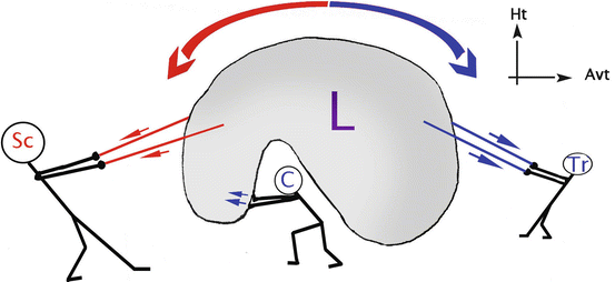
Fig. 1.12
The lunate bone is under opposite constraints; the scaphoid brings it in flexion with the scapholunate ligament, the triquetrum brings it in extension with the lunotriquetral ligament, and the capitate pressing on its posterior part brings it in extension
This allows us to understand that in case of injury of the scapholunate ligament, the lunate won’t be connected to the scaphoid and therefore will go to extension or dorsal intercalated segment instability. In case of injury of the lunotriquetral ligament, the lunate goes to flexion or ventral intercalated segment instability (Figs. 1.13 and 1.13′) [7].
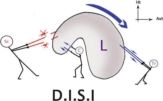
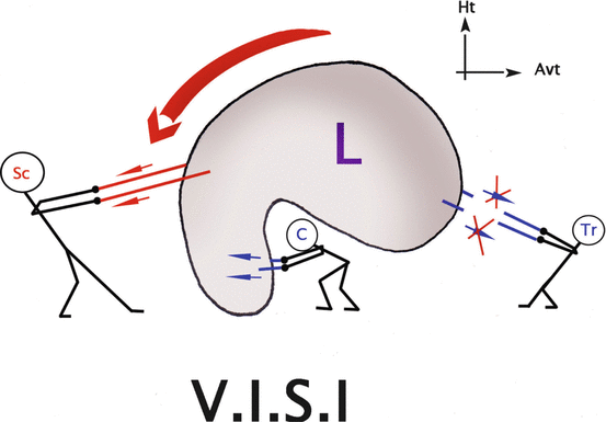
Fig. 1.13
Rupture of the scapholunate ligament, creating unbalance. The lunate tilts in DISI
Fig. 1.13′
Rupture of the lunotriquetral ligament causing a VISI
1.1.2.3 Triquetrum
It’s the most internal bone of the 1st carpal row, in the shape of a pyramid lying down with an external base and an internal top.
Its superior part is convex in every way and forms the carpal condyle with the lunate and scaphoid. The external side (2/3) of its inferior part is concave, and the internal side (1/3) is convex, to harmoniously articulate with the hamate.
1.1.2.4 Pisiform
It’s a sesamoid bone, so it isn’t really a part of the 1st carpal row. It’s a sort of “patella” for the flexor carpi ulnaris; it helps the muscle to be more efficient by improving its tendinous angle.
It articulates with the triquetrum.
1.1.3 2nd Carpal Row (Fig. 1.4)
It’s less mobile than the 1st carpal row and made from lateral to medial of:
1.1.3.1 Trapezium
It’s cubical and articulates proximally with the scaphoid and distally with the first metacarpal. Its internal side articulates with the trapezoid (superior part) and 2nd metacarpal (inferior part).
Its external face isn’t involved in any joint but is pierced with many vascular holes.
The anterior retinacular ligament, the superficial bundle of the flexor pollicis brevis and the opponens pollicis insert on its palmar side: it’s the trapezium ridge, oblique downwards and outwards.
1.1.3.2 Trapezoid
It articulates proximally with the scaphoid and distally with the 2nd metacarpal. Its external part articulates with the internal part of the trapezium and its internal part with the external part of the capitate. The deep bundle of the flexor pollicis brevis and a few fibres of the adductor pollicis insert on its palmar side.
1.1.3.3 Capitate
It’s the biggest and central carpal bone. It articulates with every other carpal bone, except for the triquetrum. Parts of the deep bundle of the flexor pollicis brevis and adductor pollicis insert on its anterior part.
1.1.3.4 Hamate
It’s prism-shaped and articulates proximally with the triquetrum and distally with the 4th and 5th metacarpals.
The flexor pollicis brevis and opponens digiti minimi insert on the hamulus on its anterior part.
1.2 Joint Anatomy and Physiology [8]
1.2.1 Radioulnar Joints
They stabilize and mobilize in order to efficiently direct the hand in pronosupination.
The amplitudes vary from one individual to the other, but it’s usually 90° of supination and 85° of pronation. The pronosupination range of motion is measured with the elbow against the trunk, with 90° of flexion to prevent the shoulder from participating to the movement.
Some authors have recently proposed the concept of “radioulnar unity”, made of the radial and proximal radioulnar joints, and a third part, the interosseous membrane [15] and the ulnar and radial diaphyses (middle lock). Each component depends on the two others.
The three joints are complementary and inseparable “locks”. If one lock is blocked, it will block the rest of the forearm. Instability in only one lock can be compensated by the two others.
Therefore, we can’t treat one of the radioulnar locks without checking the two others (Fig. 1.14).
Fig. 1.14
The three locks of the radioulnar unity
1.2.1.1 Proximal Radioulnar Joint or Proximal Lock
It’s a trochoid joint with only 1° of movement. Its congruence is maximal in an intermediary position, and it’s stabilized by a powerful ligamentous complex.
Annular Ligament (Fig. 1.15)
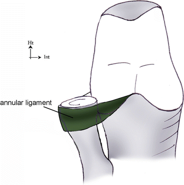
Fig. 1.15
The annular ligament surrounds the radial head. It’s a predominant element in the proximal radioulnar stability
It inserts on the anterior and posterior parts of the ulnar radial notch and surrounds the radial head with its internal face covered with cartilage. Therefore, it plays the role of a joint surface as well as a ligament.
Quadrate Ligament or Ligament of Dénucé (Fig. 1.16)
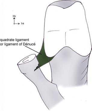
Fig. 1.16
The quadrate ligament is tensed between the radial notch of the ulna and the radial head
It inserts on the inferior part of the ulnar radial notch and ends just below the radial head, above the radial tuberosity.
Its most anterior and posterior fibres mix with the most distal fibres of the annular ligament.
It’s tensed in pronation because of its anterior fibres and in supination because of its posterior fibres.
1.2.1.2 Interosseous Membrane or Intermediate Radioulnar Joint
It’s tensed on about 10 cm between the radius and the ulna. It’s made of crossed connective fibres (collagen and elastin) that form a “network” efficiently opposed to the multidirectional constraints imposed on it:
Fibres Oriented Upwards from the Ulna Towards the Radius (Upwards and Outwards)
They form two “membranous” parts, one distal and one proximal, and a middle “ligamentous” part thicker and more resistant.
They are the most important, as their orientation allows them to transfer part of the constraints imposed on the radius towards the ulna.
In fact, 80 % of the axial constraints are transmitted on the radius through the carpus, but in the elbow, 70 % of the constraints pass through the humeroulnar joint: the load transfer distribution is inverted between the elbow and the wrist (Fig. 1.17).
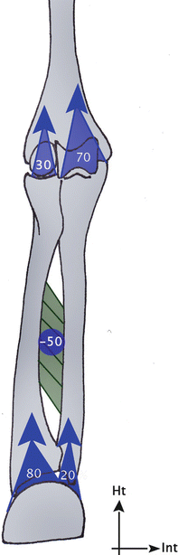
Fig. 1.17
The interosseous membrane transmits part of the longitudinal forces from the radius to the ulna, avoiding any excessive constraints on the radial head that is a fragile spongy bone
The triangular complex participates in this phenomenon at the distal level.
The interest of this load transfer is that the radial head is too fragile to bear 80 % of the constraints coming from the carpus, while the coronoid process can do it.
Recently, some studies have questioned this theory and don’t find this load transfer from the radius to the ulna.
Fibre Oriented Downwards from the Ulna Towards the Radius (Downwards and Outwards)
They form 2 individualized structures: the oblique cord and the proximal band.
The interosseous membrane is always tensed during pronosupination: the proximal part is tensed in pronation, the middle part in neutral position and the distal part in supination (Fig. 1.18).
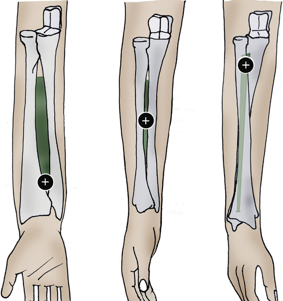
Fig. 1.18
The interosseous membrane is always tensed during pronosupination: its distal part during supination, its middle part during the intermediary position and its proximal part during pronation
The middle lock is composed by the interosseous membrane and the two bones of the forearm described in the Sect. 1.1.
1.2.1.3 Distal Radioulnar Joint or Distal Lock
It’s a trochoid joint that is neither concordant nor congruent (Fig. 1.19), which makes it particularly unstable, especially in pronation and supination. The intermediary position is the most stable one, like in the proximal radioulnar joint.
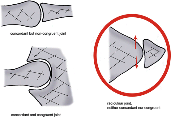
Fig. 1.19
The distal radioulnar joint is neither congruent (the bony elements don’t “fit” together), nor concordant. This leads to joint instability
It’s the radioulnar unity’s distal lock, stabilized by a powerful ligamentous complex made of:
Triangular Complex or Triangular Ligament (Fig. 1.20) [12]
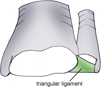
Fig. 1.20
The triangular ligament is a fibrocartilage, tensed between the radius and the ulna and bearing important constraints. It’s an important element in radioulnar and ulnocarpal stability
It’s a biconcave fibrocartilage covered with cartilage, tensed between the ulnar styloid process and the internal side of the radial epiphysis.
It’s made of an articular disc, a palmar and dorsal radioulnar ligamentous arch, a meniscus, fibres from the palmar and dorsal ulnotriquetral ligament, fibres from the ulnolunate ligament and from the extensor carpi ulnaris sheath.
It plays the role of a radioulnar ligament and the role of a meniscus hanging between the two bones of the forearm and the carpus.
Because of this, it’s under important multidirectional constraints. It opposes these constraints in different ways:
Cushioning the longitudinal constraints, as 20 % of the axial constraints of the wrist are transferred towards the ulna. It also participates to the transfer of the longitudinal forces from the radius towards the ulna, along with the interosseous membrane.
Limiting pronosupination, putting in tension the anterior fibres in supination and the posterior fibres in pronation. The most important tension is in the intermediary position, which is the most stable one.
Preventing the radius and the ulna from moving away from each other, especially when clamping (which causes the capitate to go upwards, leading to constraints on the distal radioulnar joint) (Fig. 1.21).
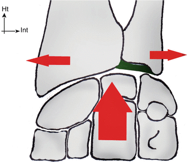
Fig. 1.21
Squeezing a ball for example, increases the longitudinal constraint on the carpus, which tends to move the ulnar apart from the radius
The triangular ligament is only vascularized in 10–40 % of its ulnar part. The middle and radial parts don’t receive any vascularization, which makes their spontaneous healing impossible.
It’s sometimes perforated in the middle, without any traumatic cause.
Radioulnar Ligaments
For some authors, they are simple capsular thickenings. They have a moderate functional role on the distal radioulnar joint. They’re located within the periphery of the triangular ligament, in a vascularized area.
1.2.1.4 Biomechanics of the Radioulnar Unity
The pronosupination biomechanics depends on the conformation of the 2 forearm bones and the functional coupling of the radioulnar joints [6].
Opposite Concavity of the Two Bones of the Forearm
It’s essential to maintain a normal pronation as it avoids the early block that would appear in pronation if the bones were straight (Fig. 1.22).
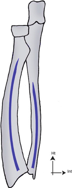
Fig. 1.22
The opposite concavity of the two bones of the forearm avoids their early blocking in pronation
Pronosupination
Pronosupination is realized according to a harmonious dynamics related to the functional coupling of the radioulnar joints.
Contrary to widespread opinion, the radius doesn’t turn around the fixed ulna: pronosupination is realized around a non-materialized evolutionary axis passing near the radial and ulnar heads.
The two bones of the forearm realize a complex movement of circumduction, in opposite directions. The humerus participates in the movement in two different ways depending on the elbow’s position (flexed or extended).
In pronation with the elbow in 90° of flexion, there’s an external rotation of the humerus inducing an external translation of the ulna, while the radius rotates on its own axis (Fig. 1.23).
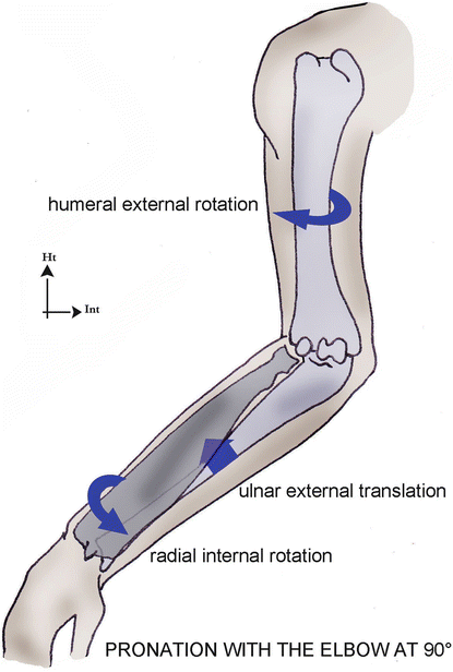
Fig. 1.23
In elbow flexion, the pronation is combined with an external rotation of the humerus
In supination with the elbow in 90° of flexion, we observe the opposite movement: internal rotation of the humerus combined with and internal translation of the ulna and a rotation of the radius in the opposite direction (Fig. 1.24).
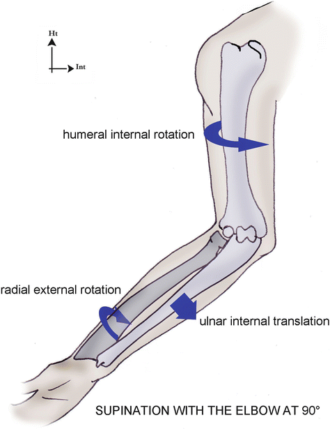
Fig. 1.24
In elbow flexion, the supination is combined with an internal rotation of the humerus
With the elbow extended, the amplitude of pronosupination is smaller, because of the tension of the humeroulnar ligaments and the loss of efficiency of the biceps and pronator teres placed in external race [4]. However, this loss is largely compensated by humeral the internal rotation in pronation and external rotation in supination, which allows the whole limb to reach 260–360° depending on the authors (Fig. 1.25).
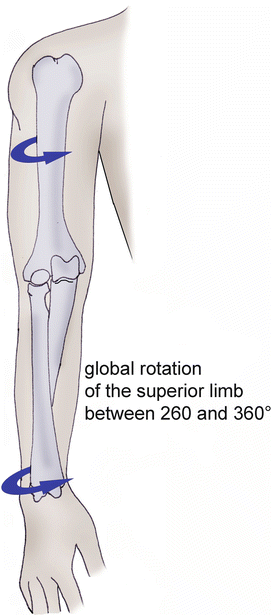
Fig. 1.25
With the elbow extended, the amplitudes of pronosupination and humeral rotation are added and allow a global rotation between 260° and 360°, depending on the authors
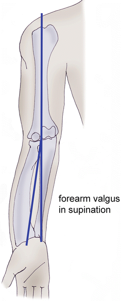
In this elbow position, the global axis of the forearm is in valgus in supination and considered as the extension of the humerus in pronation (Fig. 1.26).