Accessory bone
Prevalence (%)
Clinical significance
Differential diagnosis
Os trigonum
1–25
Synchondrotic degeneration or tear
Posterior ankle impingement syndrome
Flexor hallucis longus tendon entrapment
Shepherd’s fracture
Cedell’s fracture
Pseudoarthrosis
Accessory navicular
2–12
Synchondrotic degeneration or tear
Posterior tibial tendon dysfunction or tear
Navicular tuberosity avulsion fracture
Os sustentaculi
0.3–0.4
Synchondrotic degeneration
Painful syndrome
Isolated fracture of the sustentaculum tali
Os intermetatarseum
1.2–10
Painful syndrome
Lisfranc fracture dislocation
Os supranaviculare
1
Painful syndrome
Cortical avulsion fracture of the navicular or talar head
Os vesalianum
0.1
Painful syndrome
Avulsion fracture at the base of the fifth metatarsal
Os calcaneus secundarius
0.6–7
None
Avulsion fracture of the anterosuperior calcaneal process
Os subtibiale
0.9
None
Medial malleollus avulsion fracture
Os subfibulare
2.1
Painful syndrome
Lateral malleolus avulsion fracture
Os peroneum
9
Painful os peroneum syndrome
Bipartite os peroneum
Painful os vesalianum
Hallux sesamoid bones
Close to 100
Fracture, stress fracture, diastasis
Bipartite sesamoid
10.4 Fractures of Calcaneum
10.4.1 Epidemiology
Calcaneal fractures account for 1–2% of all fractures in adults and some 65% are intra-articular. In contrast this is a rare injury in children (below the age of 14) with the majority (70%) being extra-articular. In adolescents as the skeleton nears maturity the fracture patterns tend to resemble those of the adult with a greater proportion (60–80%) being intra-articular.
As in the adult population the majority of injuries are as a result of a fall from a height (47%), road traffic accidents (15%) and lawnmowers (13%). Up to one-third are associated with other fractures, with lower extremity fractures occurring more frequently than lumbar spine fractures. Fractures in children and adolescents also tend to be less comminuted. This may reflect the fact that these fractures in children are much lower energy injuries and the more cartilaginous immature calcaneum predisposes it to simpler fracture patterns.
10.4.2 Classification
One of the early and most widely accepted classifications of calcaneal fractures was proposed by Essex-Lopresti. This was then modified by Schmidt and Weiner for use in the paediatric population (Table 10.2).
Table 10.2
Schmidt and Weiner classification of calcaneal fractures in children
Extra-articular | 1 | A. Tuberosity or apophysis B. Sustentaculum tali C. Anterior process D. Distal inferomedial aspect E. Small avulsions off body | 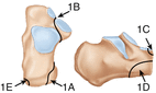 |
2 | A. Beak fracture B. Avulsion fracture of Achilles tendon insertion | 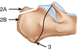 | |
3 | Linear fracture not involving subtalar joint | ||
Intra-articular | 4 | Linear fracture involving subtalar joint |  |
5 | A. Tongue type |  | |
B. Joint depression type |  | ||
Tissue loss | 6 | Significant bone loss of posterior aspect with loss of Achilles tendon insertion |  |
10.4.3 Diagnosis
Children sustaining fractures of the calcaneum are likely to have been involved in high-energy trauma. With displaced fractures, the injury to the soft tissue envelope will be directly related to the energy of the injury. The foot is often very swollen with substantial bruising and blistering. The possibility of compartment syndrome must be considered.
Standard radiographs for the calcaneum include the lateral and axial projections. Oblique views may help with identifying anterior process fractures. Bohler’s angle has been used in determining the presence of an intra-articular fracture and to quantify the degree of displacement. In adults the angle is quoted as being between 20° and 40°. In young children, the range is higher than in the adult, due to the incomplete ossification of the calcaneus. It then rapidly increases with age. By the age of 7 years the estimated mean angle is 42°. The angle tends to decline towards adult values as the child enters adolescence. Measuring the non-injured foot angle is recommended for comparative purposes, although in the case of displaced fractures the modality of choice is a CT scan.
10.4.4 Treatment
The great majority of calcaneal fractures in growing young children can be treated non-operatively due to the potential for remodelling. In the adolescent, fractures resemble the patterns seen in adults and the remodelling potential is greatly reduced. In this group, excellent results have been reported with the operative treatment of displaced intra-articular fractures.
Related posts:


Stay updated, free articles. Join our Telegram channel

Full access? Get Clinical Tree


