Sterile Instruments/Equipment
- 3.5-mm compression plates with 3.5-mm cortical screws
- 2.7-mm plates, especially for distal ulnar diaphyseal fractures
- 2.0- and 2.4-mm screws
- On-table plate bending press or hand-held bender and torquing irons
- Small pointed bone reduction clamps
- Small serrated bone reduction clamps
- K-wires and wire driver/drill
Patient Positioning
- Supine position with a radiolucent arm table.
- May use proximal arm tourniquet, if desired.
- Surgeon is usually seated in the patient’s axilla.
Surgical Approaches
- Ulna: direct approach to subcutaneous border of ulna, use interval between ECU and FCU.
- If the ECU or FCU has been traumatically disrupted, continue elevation of this muscle to avoid plating directly on the subcutaneous ulnar ridge.
- Plate may be placed on the volar surface (under FCU), on the dorsal surface (under ECU), or directly on the subcutaneous border of ulna.
- The ideal location should depend primarily on the fracture morphology.
- If the ECU or FCU has been traumatically disrupted, continue elevation of this muscle to avoid plating directly on the subcutaneous ulnar ridge.
- Radius: Volar Henry approach for exposure of the radius.
- Allows extensile exposure from proximal to distal radial shaft.
- Retract radial artery ulnarly.
- Alternatively, through sheath and bed of FCR tendon, then retract radial artery radially.
- Allows extensile exposure from proximal to distal radial shaft.
Reduction and Fixation Techniques
- For both bone forearm fractures, usually approach and reduce the fracture with the simpler pattern first.
- Restores length of the forearm anatomically.
- This facilitates anatomic reduction with the other bone and subsequently facilitates reduction of the more complex fracture.
- Restores length of the forearm anatomically.
- Multiple independent 2.0- or 2.4-mm lag screws are useful for fractures with comminution (e.g., butterfly, segmental). After interfragmentary lag screw fixation, a neutralization plate is applied spanning the area of injury.
- Usually place plates on the volar surface of the ulna (under FCU, in the flexor compartment) to avoid implant irritation as patients rest their forearms on their direct ulnar border.
- However, if either extensor carpi ulnaris or the flexor carpi ulnaris is stripped/disrupted more than the other, this muscle should be elevated preferentially.
- The plate should be placed under the elevated muscle, preserving the soft tissue attachments and hence, the blood supply of the intact muscle (Fig. 8-1).
- However, if either extensor carpi ulnaris or the flexor carpi ulnaris is stripped/disrupted more than the other, this muscle should be elevated preferentially.
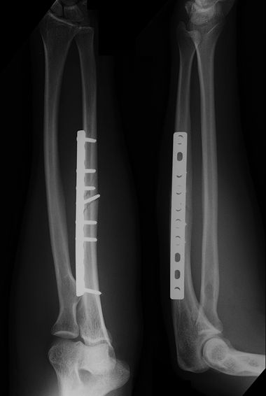
Figure 8-1. Ulnar plate placed on the flexor surface.
![]()
- For distal one-fourth ulnar fractures, consider a 2.7- or 2.4-mm compression or locking plate, especially for individuals of small stature or with osteoporosis.
- Hole spacing of the plate will allow more points of fixation in a short distal segment.
- Additionally, a 2.7-mm plate may have a better coronal plane fit than a 3.5-mm plate (Fig. 8-2).
- Hole spacing of the plate will allow more points of fixation in a short distal segment.
Figure 8-2. A segmentally comminuted ulnar fracture stabilized with two plates. A smaller plate was used for distal ulnar shaft fracture as it permitted fixation with more screws and offered a lower profile plate fit.
![]()
- Once length and stable fixation is obtained in one bone, the wound should be closed prior to performing the second approach to the other forearm bone.
- In acute trauma, generally only the skin and subcutaneous tissues are closed over the plate, to avoid compartmental syndrome.
- However, in subacute fractures, consideration may be given to closing the fascia of the FCU to the fascia of the ECU over the border of the ulna.
Related posts:

Stay updated, free articles. Join our Telegram channel
- In acute trauma, generally only the skin and subcutaneous tissues are closed over the plate, to avoid compartmental syndrome.


Full access? Get Clinical Tree


