Fixation

Sterile Instruments/Equipment
- Large/medium external fixation system
- Open compressor/distractor device
- 4.0-mm partially threaded pins for forefoot fixation
- Towel bolsters/bumps
- Dental pick
- Traingular bolster
Positioning
- Supine on a radiolucent cantilever-type table.
- Bring patient to the radiolucent (foot) end of the table.
- Place small bolster/bump under the ipsilateral hemipelvis and torso.
- Elevate the leg on soft ramp cushion to facilitate lateral imaging.
- Triangular bolster improves imaging of foot (Fig. 29-1).
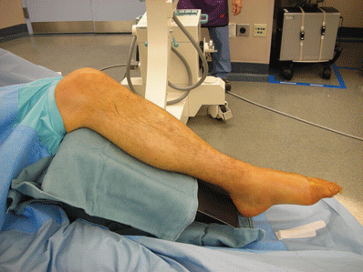
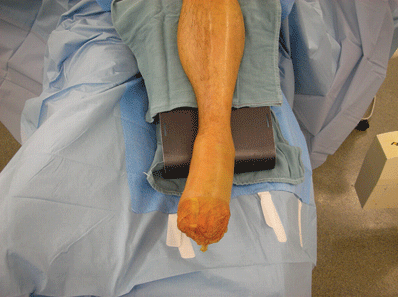
Figure 29-1. Foot position for external fixation. Note radiolucent triangle flexing hip and knee to facilitate obtaining true AP and oblique views of foot, when C-arm is in vertical position.
- With the knee fully flexed, five folded towels under the forefoot allow correct radiographic view of Lisfranc joint with AP fluoroscopy, if necessary.
Surgical Approach
Related posts:


Stay updated, free articles. Join our Telegram channel

Full access? Get Clinical Tree


