Fixation of Pediatric Femur Fractures
Patient Selection
Surgical stabilization is treatment of choice for children older than 5 years
Faster return to school
Lower overall costs than casting and traction
Several fixation options available; choice depends on patient age, size, fracture location and pattern, surgeon preference
Chapter describes techniques of submuscular plating, elastic intramedullary nailing, and lateral trochanteric-entry nailing
Submuscular Plating
Plate osteosynthesis is proven method of stabilizing pediatric fractures
Submuscular plating technique has similar advantages as plate osteosynthesis and minimizes soft-tissue disturbance
Indications
Submuscular plating recommended for patients aged 5 years to skeletal maturity with comminuted or long oblique length-unstable femur fractures
Also can be used in proximal or distal-third fractures, assuming at least two screws can be placed in proximal or distal fragments
Preoperative Imaging
AP, lateral views of femur; include ipsilateral knee and femoral neck
Evaluate for other injuries, especially to ipsilateral hip and knee
Procedure
Room Setup/Patient Positioning
Supine position on fracture table
Small (∼3%) risk of nerve palsy incidence, minimize time in traction as much as feasibly possible
Obtain provisional reduction via boot traction; evaluate length and angulation with special attention toward rotation
Place contralateral leg in extension and abduction or in well-leg holder to obtain true lateral image; perfect lateral is imperative if percutaneous techniques will be used
Can use standard radiolucent table if appropriate assistance is available
Special Instruments/Equipment/Implants
Author prefers long, narrow 4.5-mm plate; readily available, easy to contour, fits most femurs
Other options are available; choose per surgeon preference
Can use 3.5-mm plate system for smaller children
Custom pediatric implants with bowed plates are available
Can use standard or locking systems
Reserve locking plates for patients with poor bone quality or very proximal or distal fractures
Nonlocking screws help reduce fracture; if choosing locking plate system, use hybrid approach
Self-tapping screws facilitate percutaneous insertion
Plate length usually has 10 to 16 holes; need three holes for fixation proximally and three distally
Precontour plate to match lateral femoral cortex using hand or table benders
Account for greater trochanteric and distal metaphyseal flares; to maximize rigidity, plate should run length of femur
Final position of femur will match bend, so the bending should be done carefully
Surgical Technique

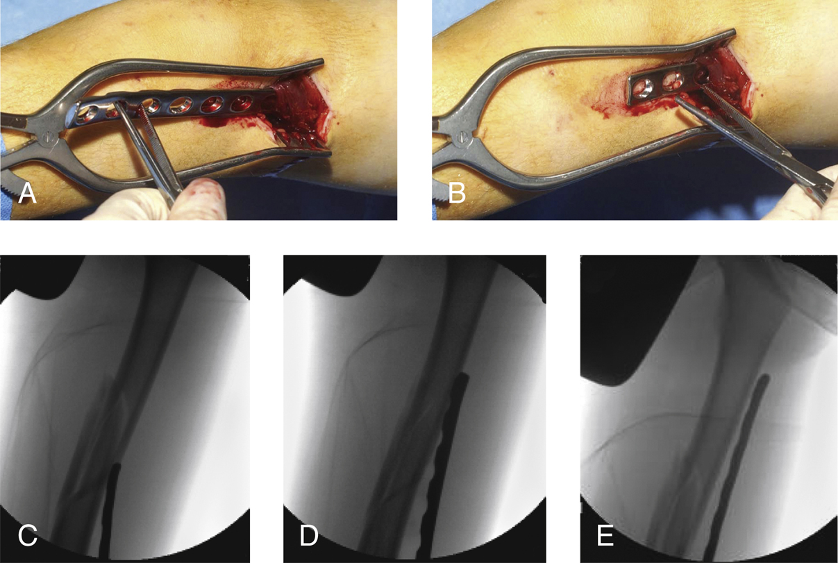
Figure 1Intraoperative photographs (Aand B) and fluoroscopic images (Cthrough E) show tunneling of the plate through the plane between the vastus lateralis and the periosteum of the lateral femur.

Figure 2Intraoperative photograph (A) and fluoroscopic images (Band C) demonstrate the reduction of the femur to the precontoured plate with percutaneous screw placement in a pediatric femur fracture.
(Panel A is reproduced with permission from Sink EL, Hedequist D, Morgan SJ, Hresko T : Results and technique of unstable pediatric femoral fractures treated with submuscular bridge plating. J Pediatr Orthop2006;26[2]:177-181.)
Make 3- to 5-cm incision proximal to the physis along lateral thigh
After incising the iliotibial fascia, identify the vastus lateralis
Carefully elevate vastus lateralis to expose periosteum
Insert plate in this interval (above the periosteum); carefully advance proximally, maintaining contact with femur (Figure 1)
Plate may be difficult to pass past fracture site; surgeon may need to pull plate back and redirect; use fluoroscopy judiciously and adjust as necessary
After plate rests in intended position, obtain AP and lateral images before placing screws
Fix plate provisionally in desired position with 2-mm Kirschner wires placed in most proximal and distal screw holes
Stay updated, free articles. Join our Telegram channel

Full access? Get Clinical Tree


