Fixation by Methyl Methacrylate
Alexandre Pagé
Andrew D. MacDowell
Paul E. Beaulé
Donald W. Howie
Polymethyl methacrylate, otherwise known as “bone cement,” has been used in the fixation of hip implants since the early 1960s. Sir John Charnley, the pioneer of modern day hip replacement, incorporated the use of cement in the development of low frictional torque hip arthroplasty.
Since then, the use of acrylic bone cement has become recognized as a highly reliable method of fixation. Cement does not act as an adhesive, as sometimes thought, but relies on an interlocking fit to provide mechanical stability at the cement–bone interface, while at the prosthesis–cement interface it achieves stability either by optimizing the fit of the implant in the cement mantle, such as in a tapered femoral stem, or by interlocking with the surface of a component such as in a cemented acetabular component.
Fixation using cement has evolved with development of improved cementing methods, changes in cement preparation, and a better understanding of how prosthesis design and surface finish can significantly improve outcome. Improved knowledge of the interrelationship between wear, periprosthetic osteolysis and aseptic loosening, and the observation that loosening also occurs in uncemented hip arthroplasty, have allayed concerns about “cement disease,” and in fact, have led some to conclude that an optimally designed cemented femoral stem will decrease the problems of periprosthetic femoral osteolysis. In this chapter, the concepts of femoral stem design and fixation, clinical results, and advances in understanding of the optimal use of cement are reviewed.
Cemented Femoral Stem Design
The design of femoral stem clearly influences the clinical result either by effects at the prosthesis–cement interface, or how the stem transfers load to cement and thereby to bone. Increasingly it is recognized that it is a combination of shape and surface finish of the stem that significantly influences long-term results. Based largely on the surface finish of the stem, and also on the way the stem interacts with the cement mantle, two main philosophies of fixation have evolved, one based around a polished stem surface, the other based on a rough stem surface, with or without adjuvant fixation features.
Polished Stems Designed to Subside
The first approach uses the highly polished surface in concert with the shape of the metal stem to optimize fit of the stem within the cement mantle and ensure that wear due to micromotion at the prosthesis–cement interface is minimized. This approach recognizes the problem of relying on a rough surface to achieve and maintain adherence between a rigid metal stem and a self-polymerizing acrylic polymer.
Of the polished stems, there are essentially three types, some of which have been successful over a number of decades. First, those modeled on the original polished Charnley stem (DePuy International Ltd., Leeds, UK), which have a rounded stem proximally with relatively rounded edges in cross section, especially medially, and a small collar; second, the collarless double-tapered stems, characterized by tapers from proximal to distal in the sagittal and frontal planes and a predominantly rectangular cross section proximally; and third, and more recently, collarless triple-tapered stems, which have an additional taper from lateral to medial. The developers of the concept of the cemented collarless double-tapered (CCDT) stems, Ling and Lee, popularized the advantages of polished stems. Initially they had proposed optimizing loading of the cement by use of a double-tapered wedge-shaped stem. Later they appreciated the importance of controlled microsubsidence of the polished stem in the cement mantle to achieve and maintain intimate contact between stem and cement and to optimize loading of the cement and, thereby, the bone (Fig. 47.1). Further, it became apparent that the collarless design and polished surface combined with the viscoelastic properties of cement could allow for stress relaxation within cement, so preserving these optimal loading conditions.
It is likely the other types of polished stems share a number of the attributes of the polished CCDT stems, and this explains the good results. However, there are some subtle differences. The small collar of the more proximally rounded stems might interfere with subsidence, so preventing intimate contact of the stem within cement, especially in suboptimal cementing conditions. Also, these designs of stem and the recent triple-tapered stems have a less-pronounced rectangular cross section proximally and so may be less resistant to posterior rotation, a now-recognized important mechanism of stem loosening.
The proposed reason for the excellent clinical results of the polished CCDT stems is that they subside within the cement mantle, usually by a few millimeters, and this mainly occurs over the first few years following insertion. Simply put, and remembering that stems are subject to considerable anteroposterior forces, this ensures the stem wedges itself
tightly into the cement mantle, thereby ensuring optimal contact between the stem and cement. Under load, axial forces are converted by the taper in the cement to radially compressive forces at the cement–bone interface. During periods of reduced load, tensile hoop strains remain within the cement, but stress relaxation can occur, with the taper maintaining the strain. Hughes et al. (1) suggest that when load is reapplied, further controlled subsidence occurs again until a new equilibrium is reached. On the other hand, should a design aim for bonding between a rough-surfaced stem and cement, stress relaxation within the construct will not occur unless the interface is disrupted.
tightly into the cement mantle, thereby ensuring optimal contact between the stem and cement. Under load, axial forces are converted by the taper in the cement to radially compressive forces at the cement–bone interface. During periods of reduced load, tensile hoop strains remain within the cement, but stress relaxation can occur, with the taper maintaining the strain. Hughes et al. (1) suggest that when load is reapplied, further controlled subsidence occurs again until a new equilibrium is reached. On the other hand, should a design aim for bonding between a rough-surfaced stem and cement, stress relaxation within the construct will not occur unless the interface is disrupted.
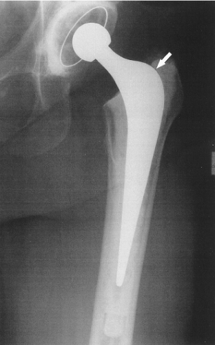 Figure 47.1. AP hip radiograph of a primary CCDT hip arthroplasty 1 year postoperatively, showing controlled subsidence (arrow). |
A proposed advantage of polished stems is that there is minimal wear of the cement and metal, should micromotion occur at the stem–cement interface. Thus wear-particle production is minimized, and any gap due to wear between the stem and cement is minimized. This, and maintenance of maximal contact by controlled subsidence, minimizes the access of wear-particle–containing joint fluid and attendant transmission of joint pressure to the femur via any defects in the cement mantle.
Anthony et al. (2) described four cases of localized endosteal bone lysis related to defects in the cement mantle in patients with otherwise radiologically well-fixed cemented matte Exeter (Stryker Howmedica Osteonics, Berkshire, UK) stems. They concluded that such defects in the cement mantle allow a route through which joint fluid could reach the cement–bone interface, subsequently leading to localized osteolysis and, ultimately, frank loosening. Fowler et al. (3) showed that localized endosteal bone osteolysis is very rare when a polished tapered stem that can subside in its cement mantle is used.
Polished CCDT stems have been very successful. Williams et al. (4) reported survivorship of 100% at 8 to 12 years for the Exeter modular stem, with the end point of revision for aseptic loosening. Yates et al. (5) reported no aseptic loosening of the CPT stem at 5 years follow-up. The 2002 Swedish National Hip Arthroplasty Register report (6) shows a survivorship of 96% for Exeter stems at 10 years, and the 2002 Danish Hip Arthroplasty Register report describes a survivorship for CPT (Zimmer Inc., Warsaw IN, USA) stems of more than 98% at 6 years (7).
Rougher Stems Designed to Bond with Cement
The other philosophy of stem design aims to achieve rigid interlock between the stem and cement and thereby nullify movement at this interface. This includes matte, grit-blasted and beaded or porous surfaces and those with indentations. A further development of this philosophy includes those stems manufactured with a precoat of cement applied to the stem with the aim of improving bonding between the stem and cement.
It is proposed that the implant–cement–bone construct acts as a “composite beam,” and the intention is to prevent movement at each of the interfaces. For this composite beam to be effective, there needs to be perfect bonding at both interfaces, with good support from the cement (8). If the construct is unable to withstand the stresses passing through it during loading, then either the cement mantle will fracture or undesirable movement will occur between implant and cement, leading to debonding at the interface. With this type of system, separation of the stem from cement, termed debonding, is thought to be an important event in the development of aseptic loosening. Harrigan and Harris, in a finite-element study of a stem designed to achieve bonding of the stem with cement, predicted a tripling of critical cement stress levels in the debonded construct compared to when bonding was present (9). Analysis of cemented stems of the rough type retrieved postmortem has demonstrated separation of these stem types from cement and fractures within the cement (10,11). Thus, debonding of this design of stem is considered a predictor of failure, but this must be distinguished from the designed subsidence of polished tapered stems, which in fact is proposed to optimize fixation.
The postmortem and other findings with rough or bonded stems has led to an emphasis on obtaining a complete cement mantle, which lessens the chance of cement cracks and failure due to mechanical reasons and also access of joint fluid to the bone of the femur, resulting in osteolysis.
Rough or Smooth Stem? How Changes in Design and Surface Finish Affect Fixation
Interestingly, during the evolution of the cemented collarless double-tapered system, there were design changes that
altered the way in which the implant behaved. When the double-tapered Exeter stem was inadvertently changed from its early polished design to a matte surface finish, the clinical results were far inferior to the early polished stems and those used for almost the last two decades.
altered the way in which the implant behaved. When the double-tapered Exeter stem was inadvertently changed from its early polished design to a matte surface finish, the clinical results were far inferior to the early polished stems and those used for almost the last two decades.
Howie et al. compared the incidence of aseptic loosening of 20 polished nonmodular tapered Exeter stems with 20 stems that were identical apart from having a matte surface finish. At 9 years, four of the matte stems had been revised for aseptic loosening, while all of the polished stems were well fixed (12). Middleton and Howie showed that matte and polished Exeter stems behave differently in the cement mantle. Polished stems subsided in the cement mantle an average of 1 mm at 2 years following insertion but did not go on to loosen up to 12 years following surgery, while early debonding at the matte stem to cement interface was associated with early loosening (13).
The Charnley stem is another prosthesis that has undergone changes in its design over the past 40 years. In a number of studies, this prosthesis has been shown to have excellent long-term results. Wroblewski et al. (14) demonstrated that even in high-demand young patients under the age of 51, the survivorship of the Charnley stem was 93% at 10 years, 74% at 20 years, and 55% at 27 years following surgery. The initial design from the early 1960s was the polished, flat-backed stem, and long-term results using this implant have been impressive, with reports of survivorship of 85% at 20 years (15). In 1976, the second-generation stem was introduced with a satin surface finish and round-backed geometrical profile. All further design changes have maintained the satin finish. Interestingly, Dall et al. (16) reported a 4- to 17-year clinical and radiologic follow-up and found an 11% incidence of stem loosening in second- and subsequent-generation Charnley prostheses but only a 3% incidence when the original design had been inserted. Schulte noted that in his series of first-generation smooth flat-backed Charnley stems, a radiographic lucent line was commonly noted superolaterally between stem and cement, indicating debonding (15). Despite this, other features of aseptic loosening did not develop, the lucent lines did not progress, and the overall survivorship of the series was 85% at 20 years despite first-generation cementing techniques. Similarly, Berry et al. (17) found that in a series of 279 consecutive smooth Charnley stems followed up for 20 years, early radiographic debonding did not have an effect on the long-term survival of the implant. However, when excessive migration with radiolucent lines of greater than 2 mm at the superolateral border of the stem was noted, there was significantly increased risk of aseptic loosening requiring revision. The latest update from Callaghan et al. (18) looked at the Charley outcome at a minimum of 35 years follow-up demonstrated a 78% survival of the Charnley total hip prosthesis at 35 years.
These findings may support Shen’s suggestion that the original smooth flat-backed design subsided in a controlled manner (8), while design changes including the satin finish and change in profile prevent this.
Crowninshield suggests that the fate of the debonded stem may depend in part on the surface roughness of the implant (19). While a rougher surface finish increases the interlock between the stem and cement, if debonding does occur, motion at the interface can produce increased amounts of abrasion with cement debris production. This would be unfavorable as it might accelerate the adverse biologic process of osteolysis and contribute to loosening.
Collis and Mohler (20) demonstrated that in a single surgeon’s extensive experience over a 27-year period, femoral stems with a rough surface were associated with higher rates of osteolysis and loosening than smooth stems. More recently, the same author compared clinical outcomes using rough and polished Iowa (Zimmer Inc., Warsaw IN, USA) stems with essentially the same geometrical profile and found that over a relatively short-term follow-up of just over 5 years, there was a significantly higher rate of revision and impending revision of the stem when a roughened grit-blasted implant was used compared to a polished implant (21). Similarly, Sporer et al. (22) found a higher rate of aseptic loosening with the rougher grit-blasted Iowa stem compared to the smoother bead-blasted version.
The issue of whether to use a cemented polished or roughened stem is hotly debated among hip surgeons. Harris argues that it is impossible to look at surface finish as an independent variable when looking at failure of cemented implants (23). He states that aseptic loosening is multifactorial in etiology and refers to several series reported in the literature of roughened stems with good results (24,25,26).
Precoating of stems with polymethyl methacrylate has been used to try to improve the bond between implant and cement, as failure at this interface is thought to be the catalyst for the development of aseptic loosening. There have been reports of high loosening rates using this technique (12,27), but other groups have demonstrated good clinical outcomes. Clohisy and Harris (28) prospectively followed up a consecutive series of 121 hip arthroplasties using a precoated stem for an average of 10 years and reported that only 1 stem needed revision for aseptic loosening. Dowd et al. (29) found that failure was more likely if there was a thin or deficient cement mantle or if the stem was not properly centralized within the canal. It may be that using precoated stems is technique dependent and works well when the stem is centralized within a good cement mantle of adequate thickness but is in danger of failure if these criteria are not met.
Stem Metallurgy
Stem metallurgy is probably as important as stem design and surface finish in determining how a cemented stem fares following insertion. Most stems inserted with cement in current practice are made of alloys of cobalt chrome or stainless steel. Historically, titanium alloys have been used with cement, with the proponents stating that the lower stiffness promotes less stress shielding of the proximal femur. This is probably beneficial in the uncemented stem scenario but has provided unique problems when combined with cement. Willert et al. revised 28 cemented Muller (Protek AG, Berne, Switzerland) straight titanium alloy stems for pain at an average of 25 months following insertion. At surgery they reported finding large amounts of titanium alloy, cement, and polyethylene debris together with evidence of crevice corrosion (11). Scalloping osteolysis was found secondary to debris particle–induced foreign body granulomas, and pain was explained by the crevice corrosion occurring in a highly acidic environment, confirmed
by pH measurements. Willert et al. concluded that titanium alloys could no longer be recommended for cementing. Other authors have had similar experiences with titanium stems. Massoud et al. (30) looked at 76 cemented titanium Capital (3M Health Care Ltd., Leicestershire, UK) stems with a mean follow-up of 26 months and found a 16% incidence of “definitely” loose stems and 10% that were “probably” loose. This, and other reports, led to the Medical Devices Agency in the United Kingdom issuing a hazard warning regarding the use of this product (31). Recently, Bowditch and Villar have suggested that the results of cemented titanium stems may not be as bad as previously reported, quoting their series of 122 arthroplasties using the cemented Howse II/Ultima (DePuy, Leeds, UK) stem with a survivorship of 97% at 7.5 years (32). However, the authors accept that these are only midterm results and that longer-term analysis is required.
by pH measurements. Willert et al. concluded that titanium alloys could no longer be recommended for cementing. Other authors have had similar experiences with titanium stems. Massoud et al. (30) looked at 76 cemented titanium Capital (3M Health Care Ltd., Leicestershire, UK) stems with a mean follow-up of 26 months and found a 16% incidence of “definitely” loose stems and 10% that were “probably” loose. This, and other reports, led to the Medical Devices Agency in the United Kingdom issuing a hazard warning regarding the use of this product (31). Recently, Bowditch and Villar have suggested that the results of cemented titanium stems may not be as bad as previously reported, quoting their series of 122 arthroplasties using the cemented Howse II/Ultima (DePuy, Leeds, UK) stem with a survivorship of 97% at 7.5 years (32). However, the authors accept that these are only midterm results and that longer-term analysis is required.
RSA Studies of Cemented Stems
Improved technology has allowed more accurate assessment of how prostheses behave following insertion. Roengten stereophotogrammetric analysis (RSA) is a sensitive tool in measuring migration of prostheses. Karrholm et al. (33) used RSA to study the migration of the femoral head in cemented Lubinus SP I (Waldemar Link, Hamburg, Germany) hip arthroplasties. At 4 to 7 years following implantation, 55 hips had not been revised, while 7 had been revised for symptomatic aseptic loosening. Interestingly, 49 of the 55 hips not revised showed some migration of the femoral head in a medial, posterior, or distal direction, or a combination thereof. This indicates that even in the clinically asymptomatic stable hip prosthesis designed to bond with cement, there is a small amount of migration of the stem. However, in the group requiring revision, RSA analysis demonstrated significantly more migration in all seven hips, the femoral head translating in a medial, posterior, and distal direction, with the total translation ranging from 4.6 to 24.4 mm. The best predictor of medium-term survivorship of the stem was found to be the amount of migration measured on the 2-year assessment, with the probability of revision being greater than 50% if the total subsidence was 1.2 mm or more.
Alfaro-Adrian et al. (34


Related posts:


Stay updated, free articles. Join our Telegram channel
Full access? Get Clinical Tree
