Intra-articular disorders
Chondral/osteochondral lesions
Subchondral cysts
Sesamoid displacement
Plica
Congenital variations
Inherited disorders
Absence
Accessory sesamoid
Symptomatic partism
Coalition
Size variations
Overuse
Avascular necrosis (see trauma)
Sesamoiditis
Chondromalacia
FHL/FHB tendinitis
Bursitis
Sesamoid trauma
Subluxation/dislocation
Stress fracture
Acute fracture
Delayed/nonunion
Avulsion fracture
Diastasis
Ligament/tendon rupture
AVN dorsal capsular tear/strain (Sand Toe)
Plantar capsular tear/strain (Turf Toe)
Metatarsal or phalangeal fracture
Subluxation/dislocation
Neurologic
Charcot neuroarthropathy
Neuritis
Arthritis
Fibromyalgia
Osteoarthritis
Crystalline deposition disease
Rheumatoid arthritis
Seronegative arthritis
Infection
Osteomyelitis
Tumor
Bone
Soft tissue
Tumor-like conditions (i.e., Ganglion)
Dermatologic
IPK/callus/porokeratosis
Epidermal inclusion cyst
Ulceration
Iatrogenic
Hallux abductus (Valgus)
Hallux adductus (Varus)
Sesamoiditis S/P Lapidus procedure
Pain dysfunction syndromes
(i.e., CRPS)
Other
1.6 Diagnostic Imaging
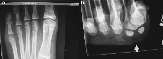
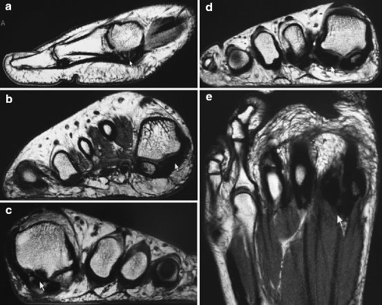
Standard weight-bearing X-rays are ordered initially and include axial sesamoid views (Fig. 1.1). Bipartite sesamoids are common and generally have a larger overall configuration than unipartite sesamoids. Proximal migration of sesamoids indicates plantar plate or flexor hallucis brevis tear and is discussed in Chap. 2. Technicium bone scans are sometimes helpful to isolate sesamoid pathology from arthritidies such as gout, rheumatoid arthritis, etc. Increased uptake on the blood flow phase indicates acute fracture while uptake only on the delayed images is associated with sesamoiditis. Computed tomography (CT) scans are helpful to show cystic changes and isolate fractures; however, magnetic resonance imaging (MRI) is becoming more common, as 3 T machines with foot coil can show fractures and avascular necrosis (Fig. 1.2).


Fig. 1.1
(a) Weight-bearing AP X-ray of a patient with a symptomatic bipartite tibial sesamoid. (b) Axial sesamoid view depicting a fractured fibular sesamoid (Photo courtesy of Amol Saxena, DPM)

Fig. 1.2
(a) Sagittal T1 MRI image of a fractured tibial sesamoid. (b) Axial MRI view showing avascular necrosis (AVN) of tibial sesamoid. (c) Axial MRI of a patient with long-standing tibial sesamoidapathy revealing adjacent metatarsal degenerative disease. (d) Axial MRI showing fibular sesamoid fracture. (e) MRI showing AVN of fibular sesamoid (Photos courtesy of Amol Saxena, DPM)
1.7 Treatment
Appropriate and specific treatment is predicated on accurate diagnosis. Types of treatment can vary significantly and is pathology and patient-type (sedentary, active, athlete) dependent. As a rule, conservative (nonsurgical) treatment is attempted in most circumstances when there is a reasonable chance it will provide a predictable result. There are exceptions to this rule though, depending on the patient type. For example, high-level amateur or professional athletes may require more definitive and efficient treatment to allow an earlier return to activity. Thus, treatment plans will need to be individualized to each patient depending on their specific expectations and circumstances.
In the case of sesamoidopathy, conservative treatments are anecdotal and have not been studied carefully. For example, in the case of an acute sesamoid fracture, recommended treatments can vary significantly from immediate weight-bearing in an accommodative orthoses or short-leg walking (SLW or below-knee) boot to prolonged non-weight-bearing cast immobilization. Because of the significant difference in treatment approaches, it is not surprising to see a high rate of delayed and nonunions in this type of injury. This disparity in method of conservative treatment can apply to most pathologies that fit under the umbrella of hallucal sesamoidopathy. The author (RTB) has coined the term “sick sesamoid”9 to apply to hallucal sesamoidopathy that is chronic in nature (>6 months), characterized by intractable pain and dysfunction, unresponsive to comprehensive conservative treatment, and pathology that has been validated by diagnostic testing. In this situation, further conservative treatment is usually futile and the patient can either live with their problem (which many patients do) or they can consider surgery. Many patients live with their problem because they are told by their health professional that sesamoid surgery does not work and the surgery will likely make them worse. This recommendation is commonly made by health professionals in general and is one of the myths that are propagated due to ignorance about this disorder.
1.8 Surgery
Surgery for hallucal sesamoidopathy can be successful and predictable even in athletic patients10 but it must be approached in a rational manner. Generically, this surgery can be considered emergent (e.g., advanced grade traumatic dorsal first MTPJ dislocation) or elective (e.g., sesamoid planing for intractable plantar skin lesion). Fortunately, most of the cases involving hallucal sesamoids are elective cases. Patients who are considering surgery are not able to live with their problem due to severity, and conservative treatment has not been successful or is not practical based on their individual situation. There are many types of surgery to consider when attempting to address hallucal sesamoidopathy (Table 1.2) and though many options are available, few have been universally accepted and recommended. Historically, surgical excision has been the mainstay but has been stigmatized by concern over postoperative complications including persistent pain, weakness, first MTPJ stiffness, and hallux deformity.11–14 This author (RTB) feels that much of the problem with surgical excision has been related to two factors: poor incision choice and not preserving the flexor hallucis brevis tendon slips.9,15 The blood supply (see sect. 1.3.2) is vulnerable to injury, and medial and plantar longitudinal central incisions have been recommended7,9 that are safer. Preservation of the FHB tendon slips is paramount to avoid hallux deformity and weakness of the flexor apparatus.16,17 Partial sesamoidectomies are preferred over total sesamoidectomies as the potential for postoperative weakness is greatly diminished.16–18 When considering surgery, there are multiple preoperative concerns to consider including patient type, concurrent foot problems, and determining whether just one or both sesamoids are pathologically involved.
Table 1.2


Surgical options
Stay updated, free articles. Join our Telegram channel

Full access? Get Clinical Tree


