Fig. 6.1
a Transverse arterial connections exist between ulnar and radial digital arteries. b There are volar to dorsal arterial connections in the hand and finger (1 Dorsal carpal arch, 2 Deep palmar arch, 3 Superficial palmar arch, a–f palmar to dorsal connections)
The Evidence: Literature Review
Many options exist for the treatment of fingertip amputations . These include primary repair, revision amputation, nonvascularized and vascularized composite grafting, skin grafting, and healing by secondary intention. Most of these well-established treatment options are surgically straightforward and are reliable choices for small defects of the fingertip.
Choosing the optimum treatment alternative depends on the goals of treatment. The primary goals of treating a fingertip defect are a functional, pain-free, sensate tip that meets the occupational and avocational needs of the patient. Secondary goals of treatment include restoration of volar pulp contour, avoidance of nail deformities, maintenance of functional length, and quick recovery.
The concept of the “reconstructive ladder” has traditionally been utilized to determine the balance between the reconstructive need of the defect and the reconstructive complexity of the surgery, with the simplest option being the preferred option (Fig. 6.2). However, this concept has been replaced with the “reconstructive elevator,” where the best surgical option according to the needs of the patient and the skill of the surgeon may be the more complex option [3]. Bennett and Choudhary stated, “Why climb a ladder when you can take the elevator?” [4]. Hence, the optimum treatment alternative and its surgical complexity will vary according to the goals of treatment and the needs of the patient (Fig. 6.3). Healing by secondary intention may be the ideal choice for straightforward defects of the fingertip . In particular, wounds with minimal bone exposure (< 3 mm), no tendon exposure, and less than 1 cm2 total surface area can be expected to heal within 4 weeks with eventual recovery of normal sensation in the majority of patients [5]. While some have expanded secondary intention to include defects > 1 cm2, this is at the expense of increased time to healing and sometimes a tender and dry scar. A patient with planing injury 2.8 × 1.8 cm in site and no bone exposed (Fig. 6.4a)is shown. In secondary intention, within hours, phagocytosis of the necrotic tissue at the end of the finger occurs , local blood vessels dilate, and an exudate forms over the wound composed of fibrin clot and inflammatory cells. Phagocytes remove necrotic debris and fibroblasts lay down bridging scar tissue (Fig. 6.4b). Peripheral epithelial cells grow inward to cover the defect. The underlying scar contracts and the defect closes (Fig. 6.4c). New nerve endings subsequently grow into the subdermal layers and sense organs repopulate the newly covered area. Other advantages of secondary intention healing include low risk of acute infection, minimal long-term stiffness, and satisfactory aesthetics [5, 6]. Long-term complications associated with secondary intention healing include 36 % cold intolerance and 26 % scar tenderness at 6 months in one study [6]. Skin grafting is another option for coverage of similar fingertip defects. Full-thickness grafts from the hypothenar eminence or a flexion crease are preferred. However, skin grafts typically have poorer sensory recovery and aesthetics than healing by secondary intention.
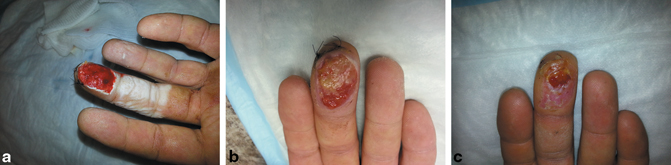
Fig. 6.2
a Reconstructive ladder. b Reconstructive elevator
Fig. 6.3
a Preoperative markings of flap for transverse distal tip defect. b Fibrous septae (white arrow) are carefully identified and released. c Flap is elevated directly off the periosteum. d Flap is advanced to cover the defect. Note the red markings identifying the preserved oblique arterial vessels. e, f Sutures are placed distally through the nail plate to secure the advanced flap. If too much tension is required to close the proximal incisions, then these may be left open to heal secondarily. g Line drawing of volar V–Y advancement flap. (Atasoy). h Line diagram of Kutler flap
Fig. 6.4
a Acute planing injury of volar pad, no bone exposed b At 2 weeks, phagocytosis of the necrotic tissue, dilation of blood vessels, and exudate formation composed of fibrant clot and inflammatory cells. c Peripheral epithelial cells move towards the center to cover the defect, which begins to close by myofibroblast wound contraction.
Revision amputation remains a popular choice for complex defects of the fingertip. In the presence of significant bone loss, tendon loss, or devascularization, revision amputation is preferred over secondary intention or skin grafting. Advantages of revision amputation include less surgical demand and quicker return to work than more complex forms of reconstruction. Complications associated with revision amputation include residual irritating nail remnant, decreased tip sensation, painful neuroma, and cold intolerance.
Nonflap options for coverage of fingertip amputations including healing by secondary intention, skin grafting, and revision amputation remain widely popular and have their established roles and indications. However, secondary intention is limited to defects of appropriate size without exposure of critical parts. Revision amputation requires shortening of the digit and is limited by poor aesthetics and risk of neuroma formation. Therefore, when secondary intention will not suffice and length preservation is desired, local and regional flaps are preferred.
Indications and Classification
When considering more complex wounds in the appropriate patient, the surgeon should employ the reconstructive elevator and consider local or regional flap coverage. These flaps can be classified as traditional local advancement flaps, staged flaps, and island flaps.
Local Advancement Flaps
An advancement flap is a segment of tissue with an intact blood supply that is advanced forward to fill an adjacent defect. The primary examples of local advancement flaps used in fingertip injuries are the V–Y advancement flaps described by Atasoy [7] and Kutler [8]. Limited fingertip injuries that involve exposed bone are ideal candidates for V–Y advancement flaps if too much bone debridement is required to allow healing by secondary intention. Patterns of amputation injury determine the most appropriate method of soft tissue advancement. In general, transverse or dorsally angulated amputations of the fingertip are suitable for a volar V–Y advancement flap as described by Atasoy and modified by others. Volarly angulated tip and apical fingertip amputations may potentially be covered by bilateral V–Y advancement flaps as described by Kutler; however, other flaps may be preferred. The advantages of V–Y advancement flaps are the low–moderate technical demand, digit and nail length preservation, and minimal donor-site morbidity.
Staged Flaps
Staged flaps involve two stages for completion of the fingertip reconstruction . The most popular staged flaps are the thenar flap and the cross-finger flap, both random pattern flaps that do not have a specific blood supply. The thenar flap and the cross-finger flap are used for volarly angulated fingertip defects, when local V–Y advancement flaps are not possible. The thenar flap was first described in 1926 by Gatewood [9]. Having undergone modification since its first description, the donor site is typically near the base of the MP joint of the thumb, which reduces the amount of finger flexion and utilizes donor skin redundancy, which reduces the degree of finger flexion and enables primary donor wound closure compared with the more proximal placement of the thenar donor site that was originally described. At the initial surgery, the flap is elevated and the injured finger is flexed to be inset into the elevated flap. After a period of time, to allow for vascular ingrowth of the recipient bed (typically 14 days), the flap is divided and inset in the volar pulp defect at a second stage. The risk inherent in the thenar flap procedure is that of recipient finger stiffness; hence, this flap is better suited for the pediatric and young adult population.
The cross-finger flap is also used in volar pulp defects with exposed bone or tendon not amenable to secondary intention healing. The dorsal soft tissue over the middle phalanx of the adjacent digit is used to cover the defect while the donor site is covered with a full-thickness skin graft. For skin graft adherence, the paratenon must be left behind on the donor digit. Like the thenar flap, the flap is divided and inset into the recipient bed after a period of time to allow for vascular ingrowth. An advantage of the cross-finger flap over the thenar flap is the ability to provide coverage to multiple traumatized fingertips in a stacked fashion. Additionally, the flap is useful in volar thumb defects where the thumb can rest comfortably against the donor index finger. Cohen and Cronin [10] described a technique for innervating the cross-finger flap to provide better sensibility by utilizing the dorsal branch of the digital nerve. Disadvantages of the cross-finger flap include finger stiffness, cold intolerance, and unacceptable cosmetic deformity of the donor site. The flap may not be suitable for elderly or female patients for these reasons.
Island Flaps
Island flaps are axial pattern flaps that maintain blood flow through a specific vascular pedicle. The idea of island flaps for fingertip coverage was not fully espoused until the principle of axial pattern blood supply to the hand and fingers was better understood. Armed with this understanding, the options for coverage of fingertip defects have flourished. Island flaps allow the surgeon to approach fingertip amputations with respect to the flap options and not the specific pattern of the defect. Island flaps may be classified as homodigital or heterodigital, antegrade or retrograde blood supply, and innervated or noninnervated. Multiple authors [11, 12] have discussed the advantages of island flaps which include:
1.
More tissue is available to completely cover large defects.
2.
Reconstruction can be completed in a single stage, which allows for early digital mobilization and reduced stiffness.
3.
Single-stage coverage minimizes the potential contamination of hardware used for fixation and tendon spacers unlike two-stage procedures.
4.
The flap is mobilized to reach the defect, not the fingertip flexed to reach the flap. This prevents immobilization in awkward positions for prolonged periods, which decreases the chance of postoperative stiffness.
5.
Island flaps have more bulk and are more similar in texture to the recipient defect than are staged flaps.
6.
Innervated island flaps allow the surgeon to provide sensate tissue to the defect with improved long-term sensibility.
7.
A greater arc of rotation or advancement can be achieved when not tethered by skin or fascial connections.
When considering the reconstructive elevator, island flaps may be considered the ideal choice for soft tissue coverage and reconstruction of fingertip defects when preservation of length and maximal function are desired and critical structures are exposed or injured.
Techniques
Volar V–Y Advancement Flap
The volar V–Y advancement flap is indicated for transverse or dorsally angulated traumatic amputations of the fingertip involving exposed distal phalanx. The advantage of this flap over revision amputation is the preservation of nail length and the potential for minimizing hook nail deformity. The blood supply of the flap is the oblique terminal branches of the digital arteries arising from the trifurcation at the distal interphalangeal joint. This is, in essence, a subcutaneous pedicle flap.
A triangular flap is designed on the volar pulp of the remaining fingertip (Fig. 6.3 shows cadaver dissection). The width of the flap distally equals the width of the defect. The flap then tapers proximally to a V at the level of the distal interphalangeal flexion crease. Leaving the apex too distal will provide an insufficient flap to advance; leaving the apex too proximal will potentially cause scar contracture at the flexion crease. The volar skin incisions are made with care taken not to incise into the deep subcutaneous tissue. Under loupe magnification, the fibrous septae are released with longitudinal blunt spreading to allow for distal advancement of the flap, while preserving the adjacent oblique vessels. A single hook may be used to grasp the distal flap to create tension and allow for visualization of any tethering septae. The flap is then elevated off the periosteum sharply to release the deep attachments of the terminal pulp fibrous septae and allow distal mobilization. Advancement of up to 1 cm can be expected with adequate release. The distal edge of the flap is typically sutured in place. Proximal sutures may be placed, but if the flap appears ischemic after tourniquet release, then the proximal incision may be left open to heal secondarily. Some have suggested spearing of the distal flap with a longitudinal k-wire as a way to inset the flap without suture and minimize tension. We have not generally recommended this.
Staged Flaps: Cross-Finger Flap and Thenar Flap
Cross-finger flaps are indicated for volar pulp defects with exposed bone or tendon. Multiple “stacked” cross-finger flaps can be used when multiple digits are involved, as is seen in industrial injuries. The blood supply to this flap is considered random.
A dorsal rectangular flap of an adjacent finger is designed over the middle phalanx to match the size of the pulp defect (Fig. 6.5). The flap is elevated off the donor digit just above the paratenon of the extensor mechanism to allow for full-thickness skin grafting of the donor defect. The flap is hinged at the midaxial line on the side of the recipient defect and inset into the defect using interrupted suture. Sensory outcomes of cross finger flaps can be improved by coapting the donor dorsal branch of the digital nerve into the recipient digital nerve stump [13]. A full-thickness skin graft is then placed over the dorsal donor defect. The patient must be splinted postoperatively to prevent shearing of the flap. At 2 weeks postoperative, the flap is divided under local anesthesia. After the second stage, an aggressive range of motion protocol should be instituted to prevent flexion contracture of the digits.