15 Femur
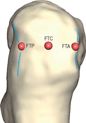
Femur – greater Trochanter (FTC, FTA, FTP)[R,L]
Landmark FTC, FTA, FTP
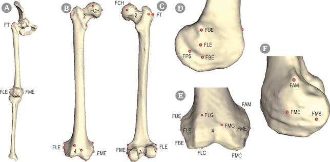
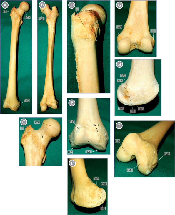
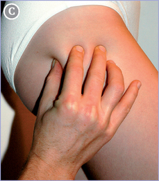
Massive quadri-angular tubercle that extends to the top of the lateral face of the femoral diaphysis (see Figs 15.1 & 15.2). It has three edges: superior, anterior and posterior.
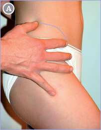
A more accurate palpation can then be performed with the three first fingers.
The FTC landmark is pinpointed by the index finger between FTP and FTA.
Femur – tubercle of the Adductor Magnus muscle(FAM)[R,L]
Landmark FAM
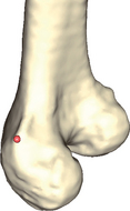
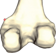
Bony protuberance situated on the superior edge of the medial condyle of the femur (see Figs 15.1 & 15.2).
![]() Subject lying: The subject is lying supine, knee flexed, with the palpator at the subject’s knee.
Subject lying: The subject is lying supine, knee flexed, with the palpator at the subject’s knee.
![]() This tendon attaches on a (usually well-developed) tubercle (FAM) above the medial epicondyle.
This tendon attaches on a (usually well-developed) tubercle (FAM) above the medial epicondyle.
Select the tubercle tip by orienting your finger pulp distally.
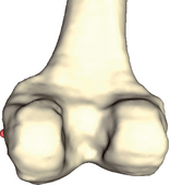
Observe the femur from a posteromedial view.
FAM is at the center of a protuberance above the medial condyle.
Turn to a posterior view to verify that the selected point is on the superior angle of the condyle.
Femur – Medial Epicondyle (FME)[R,L] ISB H|Anim
Landmark FME
This surface shows a small tubercle for the medial collateral ligament of the knee (see Figs 15.1 & 15.2).
![]() Subject lying: The subject is lying supine, knee flexed, with the palpator at the subject’s knee.
Subject lying: The subject is lying supine, knee flexed, with the palpator at the subject’s knee.
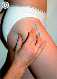
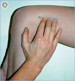
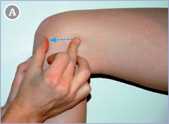
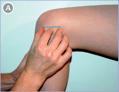
Place the middle finger on FAM (see p. 118) and the thumb on the distal edge of the medial condyle along the virtual line running distally from FAM (blue arrow).
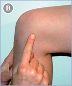
The index finger should locate a small tubercle (FME).
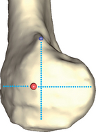
Observe the distal epiphysis from a medial view.
Find the center of the medial condyle at the intersection of the following virtual lines:
In relation to this intersection, the landmark (FME in red) to select is found slightly forwards.
Femur – Medial Sulcus (FMS)[R,L] 
Landmark FMS
Sulcus located behind and below the medial epicondyle (see Figs 15.1 & 15.2).
![]() Subject lying: The subject is lying supine, knee flexed, with the palpator at the subject’s knee.
Subject lying: The subject is lying supine, knee flexed, with the palpator at the subject’s knee.
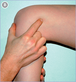
Place the middle finger on FAM (see p. 118) and the thumb on the distal edge of the medial condyle along the virtual line running distally from FAM (blue line).
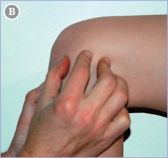
Place the tip (see p. 4) of the forefinger midway between the thumb and the middle finger.
![]() Then roll the forefinger (see finger-rolling, p. 4) backwards until its pulp center reaches the skin.
Then roll the forefinger (see finger-rolling, p. 4) backwards until its pulp center reaches the skin.
Select the point just distal to the pulp center.
Stay updated, free articles. Join our Telegram channel

Full access? Get Clinical Tree


