Femoral Revision: Classification of Femoral Defects and an Overview of Surgical Options
Craig J. Della Valle
Nicholas M. Brown
Reconstruction of the failed femoral component in revision total hip arthroplasty (THA) can be challenging. Not only can the surgical procedure itself be technically difficult, but the multiple reconstructive options available can make it confusing for the surgeon to choose among them. The purpose of this chapter is to provide a framework for selecting among the various reconstructive options available for femoral revision surgery, which are presented in more detail in the chapters that follow this overview. To this end, we classify femoral defects using the system described by Paprosky et al. (1). We have found this classification system to be the most useful as it is not only simple and reproducible, but both guides surgical treatment and yields important information regarding case difficulty and prognosis for the patient.
The first step in any revision procedure is careful preoperative planning. High-quality plain radiographs including an AP Pelvis, and AP and lateral views of the femur that include not only the end of the femoral component to be revised (or cement mantle if present) but also the femoral isthmus are required to adequately understand and classify the femoral defect and plan for its reconstruction. Further, once the plan for reconstruction has been decided upon, in general, the surgeon should always have a “backup plan” to handle the more complex problems frequently encountered intraoperatively. Finally, given the risk of intraoperative fractures in revision THA (2,3,4), even if a simple reconstruction with a shorter stem is planned, a longer stem should be available as a backup.
In general, it is our preference however to use the shortest revision stem possible (5) that will gain adequate fixation for both short-term osseointegration and long-term stability. Advantages of a shorter revision stem include avoiding the femoral bow (which greatly complicates femoral preparation and stem insertion) and preservation of bone stock for future revisions if required. Although many surgeons feel that revision femoral components must be “long,” in our experience a primary length fully porous-coated stem is adequate to handle many femoral revisions.
The Paprosky Classification
The Paprosky classification considers availability of host bone in the metaphysis and diaphysis to determine classification of the femoral defect and where fixation of the revision implant will best be obtained. It is divided into four subtypes with type 3 defects having subclassifications “A” and “B.”
Type 1
In a type 1 defect both the metaphysis and diaphysis are intact and hence the femur is essentially equivalent to a primary THA. Type 1 femurs are relatively rare, as the removal of most prior femoral components leads to loss of metaphyseal cancellous bone. This defect is typically only seen in the revision of failed resurfacing devices or if the initial femoral component was inserted without cement and lacks a surface for biologic fixation such as an Austin Moore type femoral stem (Fig. 118.1).
As previously stated, a type 1 femoral defect is essentially equivalent to a primary femoral reconstruction and the surgeon can use whichever form of reconstruction they would use for a primary THA. Once again, the unique attribute of a type 1 femur is preservation of metaphyseal cancellous bone, which allows for the use of a primary cemented femoral component if desired. However, if this form of reconstruction is selected, care should be taken to remove any membrane that may have been present around the failed femoral component to ensure an adequate cement mantle as worse than expected results have been reported following the conversion of failed hemiarthroplasties to THA (6).
Type 2
Type 2 defects are among the most commonly encountered in practice and are characterized by loss of cancellous bone
in the metaphysis and an intact diaphysis (Fig. 118.2). The metaphysis, however, is still supportive and can be relied upon for primary fixation of the revision femoral component. The loss of cancellous bone, however, precludes the use of a standard cemented stem (7) and cementless fixation is preferred. Hence, the surgeon can choose among stems that gain primary fixation in the metaphysis, as most surgeons would use for a primary cementless THA.
in the metaphysis and an intact diaphysis (Fig. 118.2). The metaphysis, however, is still supportive and can be relied upon for primary fixation of the revision femoral component. The loss of cancellous bone, however, precludes the use of a standard cemented stem (7) and cementless fixation is preferred. Hence, the surgeon can choose among stems that gain primary fixation in the metaphysis, as most surgeons would use for a primary cementless THA.
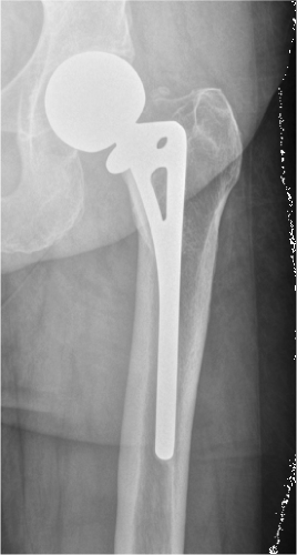 Figure 118.1. Type 1 defect with intact metaphyseal and diaphyseal bone. The prior femoral component used in the primary reconstruction in this case did not have a surface finish to allow for biologic fixation. |
However, it can be difficult for the surgeon to determine intraoperatively if the metaphysis is in fact supportive and in many ways, the use of a short diaphyseal engaging stem can be a “safer” choice for type 2 defects given the intact nature of the diaphysis with this type of defect. In other words, what looks like a type 2 femur on the preoperative x-rays may in fact be a type 3A when examined intraoperatively. Impaction grafting is another option for this type of defect, if the surgeon prefers to use a cemented femoral component, however, the previously aforementioned cementless options would in general be simpler and more economical solutions.
Type 3
In a type 3 femur, the metaphysis is damaged and no longer supportive and there is a variable amount of intact diaphysis available for distal fixation of the revision femoral component in the femoral isthmus. Type 3 femurs are divided into “A” and “B” types with 3A defects having more than 4 cm of intact femoral isthmus for distal fixation (Fig. 118.3) and 3B defects having less than 4 cm of intact femoral isthmus (Fig. 118.4


Related posts:


Stay updated, free articles. Join our Telegram channel
Full access? Get Clinical Tree
