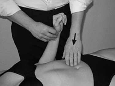
2 Fascial Treatment of the Organs according to Finet and Williame The body’s fascia consists of connective tissue and forms a continuum. In this context, we can distinguish superficial, middle, and deep fascia, but all layers are connected to each other and form a unit in the craniocaudal and anteroposterior directions. We can thus draw the following conclusion: a disturbance of the fascial dynamic anywhere in the body will, over time, cause a response in all fascia. This means that a dysfunction in a deep-lying area of the body’s fascia can be detected in superficial tissue. The fascial dynamic can be disturbed by the following: Disturbance of the fascial dynamic has consequences for the neurovegetative and hemodynamic supply of the organ: We can cite adhesions in the area of the small intestine after abdominal surgery as an example: the intestinal loops attach to the abdominal wall or to each other, and we see digestive problems or lumbar pain. The superficial fascia reacts to dysfunctions in the lower layers of the fascia with a change in tissue pull. It is the goal of visceral diagnosis of the fascia to obtain information about the lower-lying organ fascia by palpating the superficial fascial tissue pulls (induction test) and by neurovegetative reactions (hemodynamic test). By testing the superficial fascia in the abdominal wall, we identify the disturbed organ. The diaphragm is the engine of fascial movement in the abdominal organs. Migration of the organs caudally during inhalation also includes a fascial movement caudally in the abdomen. In addition to this caudal movement, the individual organ fascia carries out concomitant rotations. For the purpose of treatment, we use the respiratory movement as the mobilizing element. Normalization aims at restoring the physiologic fascial dynamic of the organ by mobilizing the superficial abdominal fascia. In this way, each organ has its specific direction for mobilization, which is discussed further in the chapters about individual organs in Section 2. Any fascial techniques of Finet and Williame that we discuss in this book consist of treatment for expiratory dysfunctions. Place your hands in the diagnostic zone of the organ and apply pressure posteriorly until you palpate the superficial fascial plane. You have reached the correct treatment plane when at a depth just before feeling the organs. To make it easier, first palpate more deeply into the abdomen until you feel the organs and then back off a little. Repeat this procedure four or five times.
Foundations
 adhesions (as a result of surgery, inflammation, or blunt trauma)
 ptoses
 viscerospasms
 parietal dysfunctions
 craniosacral dysfunctions
 The circulatory conduits penetrate the organ’s fascia to reach the organ.
 An additional vicious cycle results: if a disturbance of the fascia negatively affects the trophic state of an organ, the organ functions will be impaired, and the fascia responds in turn with an aphysiologic pull.
 Aphysiologic tissue pulls in the fascia also impair the mobility and motility of an organ. This can lead to functional impairments in the organ or parietal symptoms.
Principles of Diagnosis
Principles of Fascial Organ Treatment
Principles of the Technique for Expiratory Dysfunction
Contraindications
 acute abdomen
 carcinoma
 gallstones
 aortic aneurysm
Hemodynamic Test

Stay updated, free articles. Join our Telegram channel

Full access? Get Clinical Tree


