Extensor Mechanism Reconstruction With Marlex Mesh
Introduction
Extensor mechanism reconstruction with Marlex mesh provides durable and reliable outcomes to a catastrophic complication
Mayo Clinic experience—65 of 77 mesh reconstructions in place at 4 years, 12 patients required a revision extensor mechanism reconstruction, extensor lag improvement of 26°, with a mean extensor lag of 9°. Knee Society scores showed a significant improvement
Patient Selection
Patients undergoing or who have undergone total knee arthroplasty with an intraoperative or postoperative extensor mechanism injury
Native quadriceps or patellar tendon ruptures can be treated using this technique as well
Relative contraindication to mesh reconstruction is active infection
Preoperative (Diagnostic) Imaging
Standard knee radiographs
Closely evaluate the lateral radiograph for patella alta or baja
Less commonly, an ultrasonography or MRI may be needed
Procedure
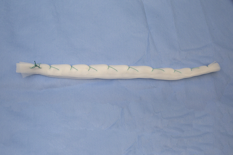
Figure 1Photograph of a 10 × 14-in sheet of Marlex mesh folded on itself eight times and then unitized with a single nonabsorbable suture.
(Reproduced with permission from the Mayo Foundation of Medical Education and Research, Rochester, MN.)
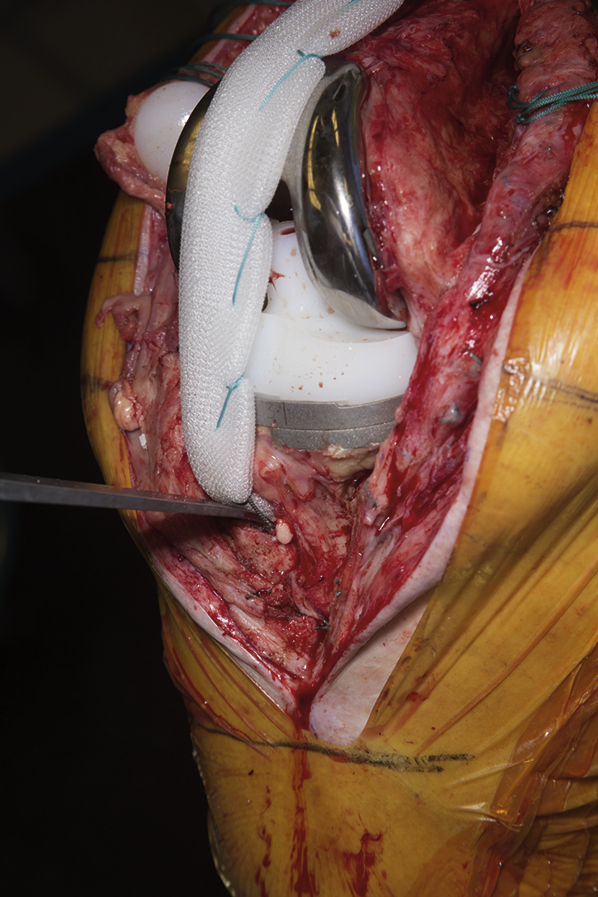
Figure 2Intraoperative photograph of the aforementioned mesh placed in the tibial trough given that the tibial component was not revised.
(Reproduced with permission from the Mayo Foundation of Medical Education and Research, Rochester, MN.)
Stay updated, free articles. Join our Telegram channel

Full access? Get Clinical Tree


