Fig. 7.1
Anterior elbow examination. The patient is seated with the elbow extended and the forearm supinated. The probe is placed on the longitudinal plane (long axis). A slight bending of the patient’s body toward the examined side makes full supination and assessment of the anterior compartment easier. Full elbow extension can be facilitated by placing a pillow under the joint
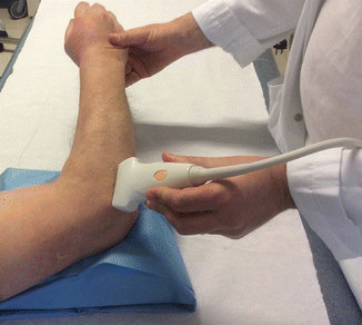
Fig. 7.2
The lateral aspect of the elbow is examined with the elbow in extension or in slight flexion. The probe is on the long axis to evaluate the common extensor tendon origin
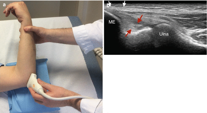
Fig. 7.3
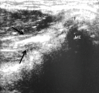
(a, b) Examination of the medial elbow. (a) The forearm is supinated and the elbow extended or slightly flexed, resting on a table with a pillow under the joint. (b) The cranial edge of the probe is placed over the medial epicondyle in the coronal plane to reveal the common flexor tendon in its long axis and the anterior bundle of the medial collateral ligament deep to this tendon. ME medial epicondyle, white arrows common flexor tendon origin; red arrows anterior bundle of the medial
7.3 US Assessment of UCL Injuries
Injuries of UCL are often injured concomitantly with the overlying common flexor-pronator mass [12]. Partial UCL tears appear as focal hypoechoic heterogeneity and ligamentous thickening [8, 11, 12]. Disruption of the UCL with widening of the ulnotrochlear joint indicates a full-thickness tear [8, 11, 12]. Dynamic valgus stress assessment allows differentiation between complete and incomplete UCL by asymmetrical widening of the ulnohumeral joint [11, 12]. Assessment of the contralateral UCL provides useful information on the normal morphology of UCL and the inherent stability of the ulnohumeral joint. This is in line with the findings during arthroscopic evaluation of widening of the ulnohumeral joint in case of a tear of the deep layer of the UCL while visually remaining intact externally [13]. US can be useful in the assessment of ulnar nerve entrapment (cubital tunnel syndrome) by demonstrating a hypoechoic thickened ulnar nerve with a cross-sectional area greater than 7.5 mm2 at the level of the medial epicondyle. A thickened, hypoechoic ulnar nerve with loss of its fascicular appearance may be seen in ulnar nerve subluxation as a result of irritation from friction during translocation. Dynamic imaging during active elbow flexion will demonstrate medial and anterior dislocation of the ulnar nerve over the medial epicondyle. Typically, the ulnar nerve will relocate within the cubital tunnel during elbow extension. Correlation with symptoms and the appearance of the contralateral nerve is important, as ulnar nerve subluxation is seen in up to 20 % of asymptomatic patients. In some patients, especially overhead athletes, the subluxation of the ulnar nerve is accompanied with a subluxation of the medial part of the triceps tendon.
The ulnar nerve, in general, subluxates over the medial epicondyle between 70° and 90° of flexion, and the triceps subluxates between 110 and 120° of flexion.
7.4 US Evaluation of Reconstructed UCL
The surgical procedure require the identification of the isometric origin of the anterior band of the MCL on the anteroinferior aspect of the medial epicondyle be identified, preserving the origin of flexor-pronator tendons. After exposure of the sublime tubercle on the medial aspect of the proximal ulna, humeral and ulnar tunnels are prepared for the passage of the tendon graft, which is then fixed using a figure-of-eight configuration or a docking technique (see Chap. 7). US is performed with the transducer placed in the long axis, with its cranial aspect over the medial epicondyle so that the hyperechoic bony contours of the medial epicondyle and ulnotrochlear articulation are seen [8]. Compared to the original anterior fibrillar band of the UCL, the graft appears as a more hyperechoic compact “cordlike” band that lay just deep to the common flexor tendon (Fig. 7.4). The US allows to follow the course of the graft from the isometric origin on the anteroinferior aspect of the medial epicondyle to the ulnar insertion close to the sublime tubercle [14]. Dynamic US is also performed to evaluate the tensile properties and the resistance of the graft with application of valgus stress on the elbow. Merolla et al. [14] in a recent research article reported good to excellent results with graft reconstruction techniques in subjects with chronic UCL insufficiency, showing similar clinical and radiographic results of allograft vs. autograft; the same authors highlights the efficacy of musculoskeletal US to evaluate the reconstructed UCL.