Glenohumeral instability is a common cause of shoulder disability. A wide spectrum of causes and presentations can make diagnosing subtle instability very difficult. This article describes clinical evaluation of the glenohumeral joint using pertinent components of the patient history, physical examination, and selective imaging to arrive at the diagnosis of glenohumeral instability in the symptomatic patient.
The glenohumeral joint is a ball-and-socket joint that achieves the greatest mobility when compared with any other joint in the human body. The overall glenohumeral to scapulothoracic motion ration is 2:1. Because of the great extremes of motion achievable by the shoulder joint, in addition to its complex anatomy that relies on a combination of bone and soft tissue structures for stability, it is not surprising that the shoulder is the most commonly dislocated joint. Although many patients who sustain an initial glenohumeral joint dislocation never experience an additional instability episode, subjective and objective recurrent instability can persist in others and may result in increased morbidity depending on the demands placed on the affected shoulder.
Despite numerous studies dedicated toward the topic, no consensus exists on what is defined as normal glenohumeral laxity. There have been many efforts to quantify glenohumeral laxity based on the amount of humeral head translation relative to the glenoid when the scapula has been stabilized. Reis and colleagues sought to quantify normal humeral head translation in cadaveric models. Mean anterior and posterior glenohumeral translation measured between 9 and 11 mm. Harryman and colleagues quantified anterior and posterior humeral head translation as 7.8 mm ± 4.0 and 7.9 mm ± 5.6, respectively, in asymptomatic volunteers using electromagnetic sensors to track humeral translation relative to the scapula. However, the difficulty in providing accurate and reproducible measuring reference points on cadaveric specimens or patients, and the great variability in humeral translation found in other studies, have raised questions regarding the validity of any of these measurements.
Historically, glenohumeral instability has been classified based on either cause or direction of instability. In general, causes of glenohumeral instability can be classified as either traumatic or atraumatic. Traumatic instability is defined as the result of an inciting event that causes subjective or objective glenohumeral subluxation or frank dislocation that is either spontaneously reduced or requires reduction by a practitioner. Atraumatic glenohumeral instability can result as the sequelae of generalized ligamentous laxity or secondary to repetitive motion; eg, activities in the overhead-throwing athlete. Glenohumeral instability can also be classified based on direction: anterior, posterior, inferior, or multidirectional. The concept of inferior and multidirectional instability was first proposed by Neer and Foster in 1980. In their report, the presence of a sulcus sign, or inferior subluxation of the humeral head relative to the glenoid with applied downward traction of the arm, with the presence of symptoms (eg, discomfort, pain, persistent episodes of dislocation, or subluxation) confirmed the presence of multidirectional instability. They identified redundancy of the glenohumeral capsule and laxity in the inferior glenohumeral ligaments as the cause for multidirectional instability.
The distinction between normal glenohumeral laxity and pathologic instability is often difficult to ascertain from history alone. The clinician must rely on the physical examination and interpretation of selective imaging to make the diagnosis of glenohumeral instability. This article describes the evaluation of glenohumeral instability, including pertinent parts of the patient history, clinical examination, and selective imaging used to arrive at the diagnosis of glenohumeral instability in the symptomatic patient.
Patient history
The examiner must obtain a thorough history from the patient regarding symptoms. Determining whether trauma to the involved extremity (eg, car accident, collision during athletic competition) preceded an initial instability event may help to determine the cause of disability to the patient. The patient may recall sustaining a frank dislocation that required manual reduction by a practitioner or a subjective feeling of instability of the shoulder “popping out,” which may represent a subluxation event. The position of the arm at the time of the instability event is a critical clue in determining the direction of instability. Patients with traumatic anterior glenohumeral instability will generally describe the instability event occurring with the arm in the abducted, externally rotated, and extended position. Patients with posterior instability, conversely, describe the arm being held in forward flexion, internal rotation, and adduction.
Many patients cannot attribute a discrete event to their symptoms. This is generally seen in patients with generalized ligamentous laxity causing pathologic instability or overhead-throwing athletes (eg, volleyball, swimming, baseball). For example, the overhead-throwing athlete may present with complaints of pain during specific phases in the throwing cycle. Pain due to anterior instability is generally experienced during the late phase of cocking. This has been suggested to be the result of repetitive stresses and attenuation of the anteroinferior, capsule-ligament complex. Posterior instability can elicit pain during the follow-through phase. Furthermore, impingement of the posterosuperior rotator cuff with the labrum can also result in pain in these patients.
Rowe and Zarins first described the “dead arm syndrome” where patients with transient anterior instability have debilitating or “paralyzing” pain or subluxation that results in brief loss of control of the affected extremity when the arm is in maximum external rotation, abduction, and extension. Jobe and colleagues further popularized this concept of instability as a source of pain in the overhead-throwing athlete. These patients may also complain of vague neurologic complaints with inferior or multidirectional instability. When neurologic symptoms exist, a history of neck pain followed by a thorough cervical spine examination must follow.
The degree of disability and the number of discrete instability events must be obtained from the history. Recurrent instability may impair the patient’s ability to perform activities of daily living (eg, carrying heavy loads, reaching for overhead objects) or may only be experienced during athletic activities (eg, pitching, striking). The ability to voluntarily dislocate the glenohumeral joint must be carefully drawn from the history, as surgical management of instability in this patient population may result in high rates of recurrence. Psychological factors may contribute to the patient voluntarily dislocating the glenohumeral joint and may necessitate psychological evaluation. However, not all voluntary glenohumeral instability patients have an underlying psychological cause. Selective activation of muscle groups can elicit instability. Patients with voluntary instability can be addressed with physical therapy focusing on biofeedback techniques.
Imaging
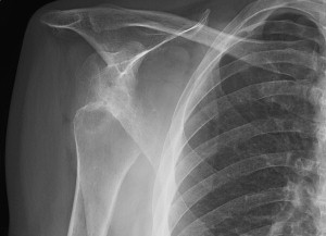
Selective imaging of the shoulder can provide critical information when assessing the patient with glenohumeral instability. Radiographic imaging of the shoulder should include; a true anteroposterior (AP) view of the glenohumeral joint in neutral, external, and internal rotation; a lateral or Y view in the scapular plane; and an axillary view ( Fig. 1 ). In the case of significant trauma, a trauma series of the affected shoulder should include a true AP view, Y view in the scapular plane, and an axillary, or a Velpeau axillary view. Hill-Sachs lesions (posterolateral impression fractures) following anterior glenohumeral dislocation can be best appreciated on the AP view in internal rotation or a notch view. The notch view, as initially described by Hall and colleagues, is performed with the patient lying supine with the cassette placed posterior to the shoulder. The hand of the affected extremity is placed on top of the head with the elbow pointing straight upward. The radiograph beam is directed 10° superior toward the head and centered over the coracoid process. Avulsion fractures and deficiency of the inferior glenoid (bony Bankart lesions) can occasionally be detected on the AP shoulder views, but are best visualized with a standard, Velpeau, or West Point axillary view. The West Point axillary view, as described by Rokous and colleagues is performed with the patient lying prone with the affected shoulder resting on a pad. The radiograph beam is aimed 25° from the horizontal plane (angled toward the table surface) and 25° toward the patient’s midline.
CT scan is a useful adjuvant test when assessing bone defects on either the humeral or the glenoid side. CT scan is particularly useful in the setting of failed shoulder stabilization surgery to determine whether bone deficiencies contribute to recurrent instability that were not adequately addressed at the index procedure. Although CT arthrography was widely used in the past, MRI and MR arthrography has become the gold standard for evaluating glenohumeral instability. Chandnani and colleagues prospectively studied the sensitivity of CT arthrography, MRI, and MR arthrography for their ability to detect glenoid labral lesions for shoulder instability. They found that MR arthrography was the most sensitive of the three techniques for detecting a detached, labral fragment and labral degeneration ( Figs. 2 and 3 ). Furthermore, MR arthrography afforded the best visualization of the inferior part of the labrum and the inferior glenohumeral ligament. Other studies have demonstrated high accuracy when using noncontrast, enhanced MR imaging for the detection of labral tears. In addition, recent studies have demonstrated the improved techniques of assessing capsular laxity in patients with recurrent anterior shoulder instability with MR imaging.
Imaging
Selective imaging of the shoulder can provide critical information when assessing the patient with glenohumeral instability. Radiographic imaging of the shoulder should include; a true anteroposterior (AP) view of the glenohumeral joint in neutral, external, and internal rotation; a lateral or Y view in the scapular plane; and an axillary view ( Fig. 1 ). In the case of significant trauma, a trauma series of the affected shoulder should include a true AP view, Y view in the scapular plane, and an axillary, or a Velpeau axillary view. Hill-Sachs lesions (posterolateral impression fractures) following anterior glenohumeral dislocation can be best appreciated on the AP view in internal rotation or a notch view. The notch view, as initially described by Hall and colleagues, is performed with the patient lying supine with the cassette placed posterior to the shoulder. The hand of the affected extremity is placed on top of the head with the elbow pointing straight upward. The radiograph beam is directed 10° superior toward the head and centered over the coracoid process. Avulsion fractures and deficiency of the inferior glenoid (bony Bankart lesions) can occasionally be detected on the AP shoulder views, but are best visualized with a standard, Velpeau, or West Point axillary view. The West Point axillary view, as described by Rokous and colleagues is performed with the patient lying prone with the affected shoulder resting on a pad. The radiograph beam is aimed 25° from the horizontal plane (angled toward the table surface) and 25° toward the patient’s midline.
CT scan is a useful adjuvant test when assessing bone defects on either the humeral or the glenoid side. CT scan is particularly useful in the setting of failed shoulder stabilization surgery to determine whether bone deficiencies contribute to recurrent instability that were not adequately addressed at the index procedure. Although CT arthrography was widely used in the past, MRI and MR arthrography has become the gold standard for evaluating glenohumeral instability. Chandnani and colleagues prospectively studied the sensitivity of CT arthrography, MRI, and MR arthrography for their ability to detect glenoid labral lesions for shoulder instability. They found that MR arthrography was the most sensitive of the three techniques for detecting a detached, labral fragment and labral degeneration ( Figs. 2 and 3 ). Furthermore, MR arthrography afforded the best visualization of the inferior part of the labrum and the inferior glenohumeral ligament. Other studies have demonstrated high accuracy when using noncontrast, enhanced MR imaging for the detection of labral tears. In addition, recent studies have demonstrated the improved techniques of assessing capsular laxity in patients with recurrent anterior shoulder instability with MR imaging.
Related posts:
 Arthroscopic Management of Anterior Instability: Pearls, Pitfalls, and Lessons Learned
Arthroscopic Bankart-Bristow-Latarjet (2B3) Procedure: How to Do It and Tricks To Make it Easier and Safe
Arthroscopic Management of Posterior Instability
Management of Failed Instability Surgery: How to Get It Right the Next Time
Arthroscopic Bankart-Bristow-Latarjet (2B3) Procedure: How to Do It and Tricks To Make it Easier and Safe
Management of Failed Instability Surgery: How to Get It Right the Next Time
Arthroscopic Management of Anterior Instability: Pearls, Pitfalls, and Lessons Learned
Arthroscopic Bankart-Bristow-Latarjet (2B3) Procedure: How to Do It and Tricks To Make it Easier and Safe
Arthroscopic Management of Posterior Instability
Management of Failed Instability Surgery: How to Get It Right the Next Time
Arthroscopic Bankart-Bristow-Latarjet (2B3) Procedure: How to Do It and Tricks To Make it Easier and Safe
Management of Failed Instability Surgery: How to Get It Right the Next Time
Stay updated, free articles. Join our Telegram channel

Full access? Get Clinical Tree


