Fig. 6.1
Glenoid shape. Sagittal oblique MR arthrogram images of a 19-year-old man with shoulder instability. (a) Normal glenoid shape exhibiting a typical pear shape. (b) Follow-up imaging obtained after an anterior shoulder dislocation shows that the inferior glenoid is narrower than the superior glenoid, the “inverted pear glenoid,” due to a large bony Bankart lesion
The damaged anterior soft tissues generally lose integrity in cases of recurrent instability because the static glenohumeral constraints become attenuated with each dislocation episode [17]. The recurrence rate after primary anterior traumatic shoulder dislocation varies widely, with reported rates up to 90–100 % in younger athletes [18]. Established risk factors that are associated with increased anterior instability recurrence rates include young age at the time of initial dislocation, associated pathologic conditions including bone loss, immobilization in internal rotation, and athletic activity [19–21].
Posterior Shoulder Dislocation
Posterior shoulder dislocations are rare events and account for less than 2 % of all shoulder dislocations [22]. It is estimated that a reverse Hill-Sachs lesion, a bony defect of the anteromedial humeral head caused by impaction against the posterior glenoid rim [17, 23], occurs in up to 86 % of posterior dislocations. Anterior cartilage damage in reverse Hill-Sachs lesions is typically more extensive than that seen in the traditional Hill-Sachs lesion [17]. Appropriate management depends on the size of the defect, extent of the disability, as well as age and activity of the patient. Given the rarity of this lesion, further discussion will focus on anterior shoulder instability.
Traumatic Glenohumeral Bone Defects
Bony defects of the humeral head and glenoid are common injuries following anterior shoulder dislocation. The incidence of osseous Bankart lesions ranges from 8 % to 90 % and Hill-Sachs lesions between 77 % and 100 %, with higher rates and sizes of defects in recurrent dislocators [24–26]. Many of these bony injuries are small and do not require surgical attention; however, there is a positive correlation between the number of recurrent dislocations and the size and extent of these osseous lesions [26]. Lesions can be grouped by the bone(s) involved: humeral head, glenoid, or “bipolar lesions” (humeral plus glenoid bone loss) [27]. With profound bone loss, patients often experience frequent dislocations with various activities of daily living, even during sleep. These positions generally include activities in which the arm is in positions of much lower degrees of abduction and external rotation than are traditionally reported during primary dislocations. A significant portion of patients with significant bone loss have failed arthroscopic soft tissue repair alone, with unaddressed bone loss attributed as a cause of failure at the time of revision surgery [3, 28]. Given that treatment options for anterior shoulder instability range from nonoperative management to arthroscopic stabilization, and even open stabilization with bony augmentation, it is imperative to accurately quantify bone loss preoperatively in order to determine the appropriate treatment plan [29].
Glenoid Lesions
There are three distinct patterns of glenoid bone loss in instability: attritional bone loss of the glenoid from prior bony Bankart injury with subsequent resorption of the bony fragment, bony Bankart with rim avulsion, and frank fracture of the glenoid. The type of injury helps to dictate the most appropriate treatment plan. In the acute setting (<3 months), fracture fragments may still be present. However, resorption of the bone fragments can occur in the months following the injury. Some rim defects occur at the time of the initial anterior dislocation. Others develop in an attritional manner, related to recurrent dislocations, as up to 90 % of patients with recurrent shoulder dislocation have at least some bony component to the Bankart lesion [30].
The size of the glenoid lesion plays a critical role in determining which lesions are significant and contribute to recurrent instability. The glenoid’s widest anteroposterior dimension is 23–30 mm with most adult patients falling between 24 and 26 mm [31]. Glenoid bone defects that are <3–4 mm (measured anteroposterior) from the anterior glenoid rim amount to between 0 % and 15 % of the total effective glenoid width and are less likely to materially affect recurrence. Defects that are greater than 6–10 mm correspond to 20–30% of total glenoid bone loss and are considered significant [30]. Sizeable glenoid bone lesions lead to decreased resistance to excessive anterior translation of the humeral head often with little applied force. Furthermore, the loss of the glenoid concavity decreases the ability of the concavity-compression mechanism in stabilizing the shoulder against anterior translation [26]. Furthermore, a narrower glenoid is more likely to “engage” a Hill-Sachs lesion in external rotation. Without addressing the bony glenoid defect, a firm glenoid socket cannot be maintained. Thus, it is largely accepted that an inverse relationship exists between the size of the glenoid defect and stability of the shoulder. Therefore, a critical component of the preoperative work-up is determining the amount of glenoid bone loss in order to dictate the appropriate type of repair: soft tissue Bankart repair alone versus glenoid restoration or augmentation with a Bristow or Laterjet reconstruction [32].
Hill-Sachs Lesions
As stated, compression fractures of the humeral head are very common injuries. The challenge lies in predicting which humeral lesions contribute to recurrent instability. The size, orientation, and location of the lesion should be determined on preoperative imaging and during arthroscopy, as all have implications for treatment. Assessments are based on the size of the lesion (length and depth), location along the posterolateral aspect of the humeral head, and the percent involvement of the 180° articular arc [17]. There is controversy regarding the threshold size and precise location of the defect that will materially contribute to instability; however, large Hill-Sachs are considered a risk factor for postoperative recurrence because larger humeral lesions more freely engage the glenoid rim [25]. Small Hill-Sachs lesions, less than 20 % of the humeral head curvature, are generally not considered significant sources of recurrent instability [33], though some authors report lesions as little as 12.5 % of the humeral head may prove consequential to shoulder instability [34]. Regardless, most authors agree that Hill-Sachs lesions greater than 40 % of the humeral head curvature are significant enough to warrant surgical treatment [2, 20, 21]. Lesions that are 20–40 % may be significant depending on their location, orientation, engagement, and coexistence with a glenoid bone lesion. The combination of a Hill-Sachs lesion and glenoid defect substantially reduces the normal arc of shoulder movement [33]. If a Hill-Sachs lesion is present, a dynamic examination should be performed at arthroscopy in which the shoulder is brought through full range of motion in order to discern “engagement” of the humeral head defect with the anterior glenoid rim [3].
The Engaging Lesion
Burkart and De Beer [35] were the first to report that one of the factors responsible for failure of arthroscopic soft tissue stabilization was traumatic bone deficiency, introducing the concept of “significant bone loss of the humeral head and glenoid.” Significant bone loss of the glenoid was defined at arthroscopy if the glenoid had the appearance of an inverted pear. A significant humeral head bone defect was defined at arthroscopy as an “engaging” Hill-Sachs lesion: a lesion that presents parallel to the anterior glenoid when the shoulder is placed in a functional position of abduction and external rotation. The location of the defect allows the lesion to engage or “hook” the corner of the anterior glenoid due to an articular-arc deficit (Fig. 6.2). Conversely, a nonengaging Hill-Sachs lesion was defined as a defect that is presented in a nonparallel angle to the anterior glenoid in a functional position (abducted and externally rotated) or one in which engagement occurs in a nonfunctional position of shoulder extension or of low shoulder abduction (<70° abduction). This study emphasized the role of arthroscopy as a dynamic diagnostic tool. The authors advocated for not only repair of the soft tissue Bankart lesion but also an operative measure to address significant bony lesions in order to prevent the Hill-Sachs lesion from engaging. In addition, the authors confirmed the higher likelihood of substantial glenoid bone loss in patients with recurrent dislocations. Though the sizes of the lesions were not defined, other studies have shown that larger volume lesions are more strongly associated with recurrent shoulder dislocation [36, 37].
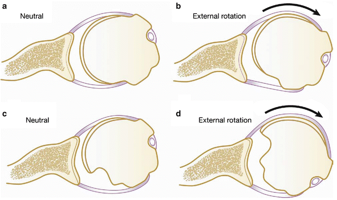
Fig. 6.2
Engaging lesion. (a), (b) show the normal relationship of the humeral head with respect to the glenoid when the shoulder is abducted and externally rotated. (c), (d) depict a large Hill-Sachs lesion. When the shoulder is abducted and externally rotated, the humeral head defect can engage or hook the inferior rim of the glenoid due to an articular-arc deficit [Reprinted with permission from Burkhart SS, Lo IKY, Brady PC. A Cowboy’s guide advanced shoulder arthroscopy (ed 1). Philadelphia: Lippincott, Williams & Wilkins, 2006]
Itoi et al. [38] supported the importance of bone loss in a three-dimensional (3D) CT imaging study, reporting that a glenoid defect with a width that is at least 21 % of the total glenoid length may materially affect recurrence after instability surgery. As a result, restoring the width of the glenoid may be beneficial in limiting recurrence. Other authors focused on the size of the Hill-Sachs lesions as a predictor of instability. Rowe et al. [39] classified Hill-Sachs lesions into three sizes and demonstrated that there was an increasing rate of recurrent dislocation with larger bone defects. Several authors have reported positive results of small case series in which patients with Hill-Sachs lesions greater than 20 % of the humeral head underwent reconstruction with allograft [40–42]. The confusion and continued debate in the literature regarding which bony lesions truly need to be addressed operatively laid the ground work for the critically important concept of the “glenoid track.”
The Glenoid Track
Yamamoto et al. [24] introduced the concept of the “glenoid track ” to determine which Hill-Sachs lesions have the potential to engage with the glenoid. Using cadaveric shoulders and 3D CT imaging, the authors demonstrated that as the arm is elevated, the glenoid contact area with the humeral head traveled from the inferomedial to the superolateral portion of the articular surface of the posterior humeral head, creating a zone of contact which the authors referred to as the “glenoid track.” If the Hill-Sachs lesion falls lateral to the glenoid track, there is minimal likelihood of engagement. On the contrary, if the margin of a Hill-Sachs lesion falls medial to the glenoid track, there is significant risk that the humeral head will override the glenoid rim and engage. The authors computed that the width of the glenoid track (medial margin of the contact area to the medial margin of the rotator cuff attachment) is 84 % of the width of the glenoid. With glenoid bone loss, as the width of the glenoid track decreases, the probability that the Hill-Sachs lesion will fall medial to the “track” increases and thus “engagement” is more likely to occur. This concept has been very useful because the authors introduced the notion that not only the size of the Hill-Sachs lesion but also its location impacts engagement. Furthermore, the glenoid track concept highlights the interdependence of both glenoid and humeral bone loss in predicting recurrence. The width of the glenoid track and the size and location of the Hill-Sachs lesion are closely linked factors that need to be considered when addressing anterior instability.
All bipolar (glenoid and humerus) lesions can be considered “engaging,” as the initial insult led to the glenoid rim-humeral contact necessary for the formation of bone loss. While Burkhart and DeBeer initially applied the term “engagement” to refer to the abduction and external rotation position, in truth, bone loss can only occur when the glenoid rim and humeral head contact. If the initial insult is repeated, bipolar lesions become increasingly more likely to engage. While dynamic intraoperative arthroscopic assessment of engagement is performed during Bankart repair, this diagnostic technique may lead to overestimation of whether a Hill-Sachs engages because concomitant ligament insufficiency is commonly present. Such laxity may permit excessive anterior translation or external rotation of the humeral head and facilitate engagement that would not be present when ligamentous integrity was present [27]. The prevalence of engaging Hill-Sachs lesions has been reported to be between 1.5 % [23] and 33 % [43]; however, the term “engagement” needs to be more precisely defined.
Further work by Kurokawa et al. using the glenoid track concept [25] sought to determine the prevalence of Hill-Sachs lesions that need to be addressed surgically. Using CT images, the authors observed that 7 % of 100 shoulders which demonstrated recurrent anterior instability had Hill-Sachs lesions that extended medially over the glenoid track. The authors divided the seven engaging lesions into two subtypes: a wide and large Hill-Sachs lesion (three cases) and a narrow but medially located Hill-Sachs lesion (four cases). This study supports the notion that not only the size of the Hill-Sachs lesion predicts engagement but also the medial extent of the lesion. All seven engaging Hill-Sachs lesions also demonstrated large (20 %) glenoid defects, supporting the increased risk of engagement from decreased width of the glenoid track secondary to the bony glenoid defect itself.
Arthroscopic Assessment of Glenoid Bone Loss
Glenoid bone loss can be measured directly arthroscopically by referencing the glenoid bare area, as first described by Burkhart et al. [44]. The etiology and development of the bare area are unclear, but it is thought that the bare area is a region of attenuated cartilage that arises due to repetitive stress loading over the subchondral tubercle of Assaki [45]. The normal glenoid is shaped like a pear; Burkhart measured the distance from the bare area to the anterior, posterior, and inferior glenoid margins in 56 patients without instability and found that the bare area was approximately equidistant from the anterior, posterior, and inferior margins. Since the majority of bone loss in anterior instability is anterior and inferior, the amount of bone loss can be calculated by factoring in the distance from the bare area to the posterior margin and assuming this distance is roughly the same from the bare area to the anterior rim in a normal shoulder.
Subsequent studies have questioned the utility of the bare area as a consistent reference point in glenoid bone loss determination. The main criticism of this technique is that the bare area is absent or eccentrically located in a large percentage of patients [31, 46]. Barcia et al. [47] recently showed that in a series of 52 patients undergoing arthroscopy without a diagnosis of instability, the bare area was present in only 48 % of patients. In patients in whom the bare area was found, it was centrally located in only 37 % [47]. Thus, an anterior bare area would lead to overestimation of bone loss and potential unnecessary glenoid augmentation procedures. Conversely, a posterior bare area would lead to underestimation of bone loss, and those patients may not undergo glenoid augmentation when it is indicated. Due to the limitation in intraoperative arthroscopic measurements, preoperative imaging is therefore required to accurately and reliably calculate glenoid bone loss.
Imaging Techniques
Radiographic Evaluation
The initial evaluation of shoulder instability should begin with a conventional radiograph series: a true anteroposterior (Grashey) view, internal and external rotation views, a scapular Y-view, an axillary lateral view, and an apical oblique (Garth) view [3]. These projections demonstrate the relationship of the humeral head to the glenoid and allow for assessment of bone defects, joint space narrowing, and osteophytes. Each projection plane offers unique information in the assessment of instability. The Garth view is optimal for evaluating bony Bankart fractures and Hill-Sachs lesions [30]. Additional views such as the West Point axillary and Stryker notch views may provide additional detail about the size of the bony Bankart and Hill-Sachs lesions, respectively. The West Point axillary view is designed to assess defects within the anterior-inferior glenoid rim [26]. The Stryker notch view, obtained by placing the hand on top of the head and X-ray beam angled 10° cephalad, can be used to evaluate the presence, size, and orientation of the Hill-Sachs lesion, as the internal rotation of the humeral head brings the defect into direct view [17]. Radiographic measurements are typically performed by measuring the defect or notch width and the depth with respect to a tangent line. Cross-sectional imaging has become increasingly popular in assessment, and its usefulness is discussed later in this chapter.
In many cases, additional cross-sectional imaging is required for a complete evaluation, as half of all bony lesions may be missed by conventional radiographs, and it is difficult to discern lesion size on plain films alone [48]. Therefore, those patients with a negative plain film evaluation, but a history and physical examination concerning for bony defects, should undergo additional cross-sectional imaging with either CT, MRI, or both, in order to accurately quantify bone loss. Arthrography should be used to increase the sensitivities of these examinations unless there is a contraindication to joint injection. Patients with evidence of glenoid boss loss on conventional radiographs will still benefit from additional cross-sectional imaging in order to better characterize the size, location, and extent of the bony lesion, as well as to evaluate for concomitant soft tissue injury such as rotator cuff tears and bicep tendon injuries, labrocartilaginous injuries, and capsuloligamentous disruptions [3].
Computed Tomography
CT imaging combined with 3D reformatting is a superior, noninvasive option for evaluating bone loss due to superb contrast between bone and soft tissues. The usefulness of CT scanning to define the morphological characteristics of glenoid defects and Hill-Sachs lesions and quantify the amount of bone loss has been well documented [2, 17, 49, 50]. Reasonable indications for CT scanning include multiple dislocations, bilateral shoulder dislocations, failed stabilization procedures, dislocations after trivial trauma with little or no provocation, radiographs or MRI demonstrating significant bone loss, and instability in midranges of motion [30].
As noted above, accurate assessment of glenohumeral bone loss is imperative in guiding surgical management. There are various methods to assess the size of the bony glenoid defect: defect length, width to length ratio, glenoid index (defect width/circle diameter), Pico method (defect area/circle area), and glenoid arc angle [27, 29, 51]. These measurements can all be performed after the acquisition of a standard CT scan of the shoulder. A standard shoulder CT protocol is performed with acquisition of both shoulders simultaneously in the axial plane, scanning from the superior aspect of the acromion to the inferior aspect of the glenoid fossa. The arm should be kept down in a neutral position. General parameters include field of view (FOV) 48 cm, pitch 0.9, collimation 1 mm, 120 kV, 200 mA (or dose modulation) with 1 mm image reconstruction. Image reformations are generated in the coronal oblique and sagittal oblique (with the glenoid en face) planes. In addition, 3D reconstructions should be created. After digital subtraction of the humeral head from the glenohumeral complex, the scapula and glenoid fossa can be optimally visualized as they are no longer obscured by the humerus, and glenoid bone loss can be precisely quantified. Due to the convention of CT imaging, the contralateral shoulder is imaged at no additional cost and can be used for comparison. In healthy subjects, there has been no significant difference in right-sided and left-sided glenoid measurements [49]. It is important to stress that thresholds for “critical” bone loss need to be determined specifically for the measurement technique being used.
Related posts:


Stay updated, free articles. Join our Telegram channel

Full access? Get Clinical Tree


