Preoperative Evaluation and Testing for Arthroscopy
Keywords
• Arthroscopy • Ankle surgery • Preoperative planning • Ankle imaging
Indications
Currently, there are no absolute contraindications for arthroscopy of the ankle.1 However, there are some relative contraindications, such as patients with compromised circulation or patients with comorbid medical issues.
Glazebrook and colleagues1 reported “that the basis for arthroscopy for indications such as ankle instability, septic arthritis, arthrofibrosis, removal of loose bodies or ankle arthritis in the absence of bony impingement lacked sufficient evidence based support.” Conversely, they reported that, “preparative ankle arthrodesis approaches, osteochondral lesion repairs and treatment of ankle impingement syndromes were fairly supported as indications.” For the purpose of simplification, relative indications for ankle arthroscopy can be divided into 3 distinct surgical categories based on the desired final outcome for the procedure:
Indications for an ankle arthroscopic survey include lavage for septic joint with survey, syndesmotic analysis, preemptive assessment of joint before an intended open repair, assessment of poorly placed internal or external fixation hardware, and arthroscopic biopsy. With respect to arthroscopic survey, the scope of the procedure is relatively narrow, as one would expect with any operative survey. Surveys may be performed after an examination under anesthesia with mortise and Broden’s views of the ankle under image intensification before a formal repair of the lateral ligament and retinacular structures or “Brostrom” (modified or true) repair for ankle joint instability. An arthroscopic survey may also be beneficial as a diagnostic tool when infection is suspected. “Ankle joint surveys performed to inspect and treat septic ankle joints have also been successful as a treatment modality though there is a paucity of literature to support this technique.”1,2 The success of this approach may be directly related to the physiologic lavage and reduction of a pathologic microorganism count more so than the topical introduction of antibiotic-rich saline.
Another parameter in the surgical decision-making process as to whether an open repair versus an arthroscopic procedure is better indicated can be made on realization of the constraints of an arthroscopic approach to the ankle joint. Studies have shown that patients with bony or soft tissue impingements tend to do better with smaller focal impingements and a lack of significant osteoarthritis.1,3,4 This consideration is an important one if solely for the purpose of open treatment consent and appropriate instrumentation being available at time of surgery.
If there is definitive presence of an osteochondral defect (OCD),1,5 a reparative arthroscopic approach may be attempted to reduce the defect and relieve pain.1,4,6 However, careful consideration should be given to the location of the defect. The location of the lesion may play a significant role in the postoperative outcome of the repair. A talar dome defect or tibial plafond defect may respond better to arthroscopic repair versus a shoulder defect of the talus or a medial/lateral gutter defect. Kelberine and Frank compared anterolateral OCD repair versus posteromedial repair and reported that the anterolateral group had significantly better results (89%) versus the posteromedial study group (63%) with regard to patient improvement and overall satisfaction.5 This is a statistically significant finding when transposed against the long-accepted notion that anterolateral OCD repair should not be attempted owing to the lack of improvement and the location of the lesion. It should also be noted that posteromedial defects are commonly associated with plantarflexion/inversion sprains, whereas anterolateral lesions are more commonly a result of a dorsiflexion/inversion injury. These 2 common defects also differ in their presentations from an arthroscopic appearance. Anterolateral lesions are typically “wafer” shaped and relatively superficial, whereas posteromedial lesions are typically more “cup” shaped, indicating a deeper injury. In addition, the proximity of anterolateral OCDs to structures that may be contributing pain sources may also add to the higher postoperative scores by easier access to these structures during the defect repair.5,6 Conversely, posteromedial lesions may be difficult to access at the time of surgery and may require malleolar osteotomy or a posteromedial approach depending on the size of the lesion and its location. For example, an anterolateral soft tissue impingement by traumatic thickening of Bassett’s ligament can be repaired concomitantly during the repair of an anterolateral OCD.
Relatively new indications for ankle arthroscopy are always emerging that include joint debridement for arthrodesis, management of septic joints, and aid in fracture reduction. Thermocautery or “capsular shrinkage procedures,” intra-articular fracture reduction, and ankle arthrodesis procedures are relatively new and are gaining support as indications for ankle arthroscopy. In cases of arthroscopic thermocautery for ankle joint instability, Berlet and colleagues7 and Hyer and Vancourt8 reported good results with lateral ankle stability using thermocautery with lateral ankle gutter debridement.
Intra-articular open reduction of fractures via ankle arthroscopy has also become more popular in recent years both intraoperatively and postoperatively.1,3,9 During an open reduction of an intra-articular fracture of the ankle joint complex (tibia, talus, and fibula), arthroscopy can be used to verify anatomic reduction of the joint surfaces with debridement of any cartilage defects that may be present. Postoperatively, if painful hardware becomes an issue, arthroscopy also can be performed with hardware removal as an adjunctive procedure to enhance postoperative results in the presence of minimal posttraumatic arthritis.3
Surgical ablative arthroscopy is indicated when the procedure is used as a surgical “means to an end” to a more comprehensive procedure, such as joint preparation for ankle joint arthrodesis. The increased usage of this technique to perform a “minimally invasive” arthrodesis procedure versus a traditional open ankle arthrodesis has been fairly well supported by current literature.1,10–12 Good results have been demonstrated with use of this technique.1,10,11 This minimal approach may be gaining acceptance because of the reduction in tourniquet time and wound morbidity associated with traditional open procedures.1
Patient history and the physical examination
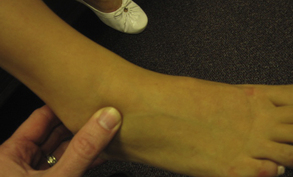
Examiners should take a systematic, “around-the-world” approach to examining the ankle joint. This approach is performed at the farthest point from the suspected pain foci and progresses to the foci in both directions in a circumferential matter. When performed repeatedly, the examiner will develop a unique proficiency for recognizing certain ankle pathologies. All of the bony and soft tissue structures that pass the ankle joint should be palpated as well as palpation of the joint line and its corresponding landmarks. By working in a continuous manner about the ankle joint, the examiner will be able to eliminate redundancies (referred pain) in presentation as well as adding to the efficiency of the examination. Patients should be sitting up on an examination chair or table with the lower extremities in open chain attitude (heels free) for an accurate physical examination. Some examples of palpation tests are shown in Figs. 1 to 6.


Stay updated, free articles. Join our Telegram channel

Full access? Get Clinical Tree


