Epidemiology, Risk, and Risk Factors


Epidemiology, Risk, and Risk Factors


Introduction

Osteoporosis is defined in terms of bone quality, bone mass, bone architecture, and fracture risk. The epidemiology of osteoporosis therefore relates to all these and to their outcomes. Clinically, osteoporosis is recognized by the occurrence of fractures following low-energy trauma, the best documented of these being hip, vertebral, and distal forearm fractures. The definition has been previously considered (Table 1.1).

The incidence of osteoporosis is best measured indirectly as the incidence of fractures that are attributable to the condition. Prevalence is best measured by the frequency of reduced bone mineral density (BMD) or numbers of those with vertebral deformity. The risk of sustaining a fracture related to osteoporosis can be considered for the future lifetime from the age that the risk increases – that is from 50 years. Alternatively, risk can be considered as the probability of sustaining a fracture during a meaningful period and 10 years is considered more appropriate when deciding whether or not to intervene to reduce that risk.


Epidemiology of osteoporosis

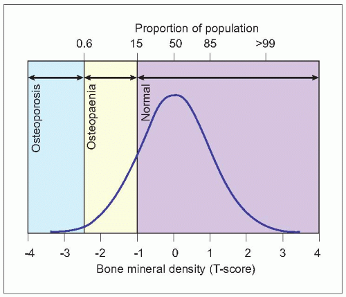
There is a normal distribution of bone density across the population for any age and either sex (2.1). Bone strength declines and the risk of fracture increases with reduced bone density. Normal bone density is defined as being -1 standard deviation or greater than the mean at 30-40 years (peak bone mass). Bone density between -1 SD and -2.5 SD of peak bone mass (T-score) has been defined by the WHO as osteopenia, and equal or below 2.5 SD of peak bone mass as osteoporosis1.

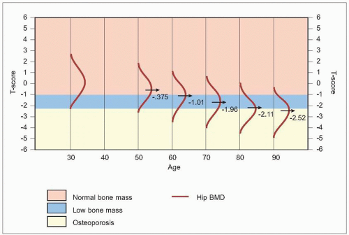
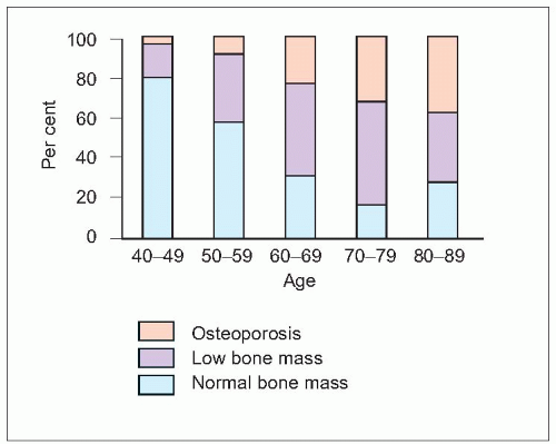
As bone density declines with ageing, the number at any age with osteoporosis will increase (2.2)2. It is estimated that 54% of postmenopausal white women in the northern USA have osteopenia, and a further 30% have osteoporosis in at least one skeletal site. In the UK, it is estimated that around 23% of women aged 50 years or more have osteoporosis as defined by WHO. For the diagnosis of osteoporosis, it has been recommended by the International Osteoporosis Foundation to measure BMD by dual energy X-ray absorptiometry (DXA) at the hip, and by this definition the general prevalence of osteoporosis rises from 5% in women at the age of 50 years to 50% at the age of 85, and in men the comparable figures are 2.4% and 20%3. The dramatic increase from midlife into older age in the percent of women who have osteoporosis in Sweden is demonstrated in Figure 2.3. This is set against the trend of longevity with women aged 80 having a life expectancy of over 8 years.


2.1 Distribution of bone density in healthy women aged 30-40 years. (Adapted from WHO (2003). Prevention and Management of Osteoporosis. WHO Technical Report Series No. 921. World Health Organization, Geneva.)


2.2 The distribution of bone density at increasing ages. (Adapted from Kanis J, et al. (1994). The diagnosis of osteoporosis. J Bone Mineral Res 9(8):1137-1141.)


Epidemiology of fracture

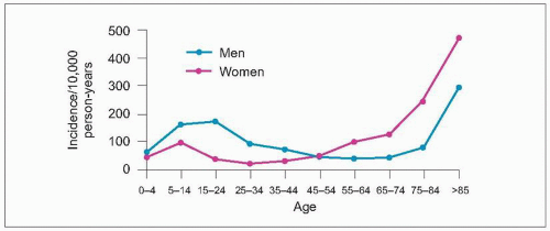
Fractures occur most frequently in adolescents and young adults and, in later life, increase from age 50 years (2.4). At 75 years, over 40% of women will have sustained a fracture4. In adolescents and young adults they are more frequent in males than females when they are usually associated with major trauma such as road traffic accidents and sports injuries. The sites of fracture are usually long bones, most commonly the forearm5. In later life, fractures are more common in females than males and follow low-energy trauma, most commonly falls. The majority of these fractures in those aged over 50 years are the result of underlying osteoporosis.

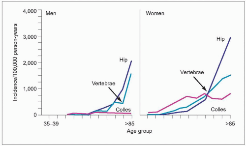
The fractures that are most strongly associated with age and osteoporosis are of the distal forearm (Colles’ fracture), the proximal femur, and of the vertebrae (2.5). These sites are predominantly trabecular bone. The incidence rates of proximal humeral, rib, clavicle, and scapula also rise with age and are greater in women than in men. About 80% of proximal humeral fractures occur in individuals aged 35 years and over, three-quarters occurring in women.


2.3 The percentage of women in Sweden with normal bone mass, low bone mass, and osteoporosis in the hip at increasing ages. (Adapted from SBU (1995).

Measurement of bone mineral density. SBU Report 127. Swedish Council of Technology Assessment in Health Care, Stockholm.)


2.4 Incidence of fractures in the UK. (Adapted from Donaldson LJ, et al. (1990). Incidence of fractures in a geographically defined population. J Epidemiol Community Health 44(3):241-245.)


2.5 Incidence of the major age-related fractures in men and women. (Adapted from Cooper C, Melton LJ (1992). Epidemiology of osteoporosis. Trends Endocrinol Metab 314:224-229.)


Vertebral fractures

The incidence and prevalence of radiological vertebral deformities increase with age. One in eight men and women over 50 years in Europe are estimated as having vertebral deformity from a large epidemiological study conducted across 18 European countries6. Vertebral deformity is describing a radiological finding (see Chapter 1) and it is not possible to say with certainty when and how it arose. This term is used in epidemiological studies. If it appears that the deformity is related to a fracture, then that term is used.

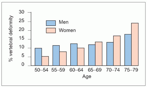
Vertebral deformities are more prevalent in males than females aged 50-59 years and there is a less dramatic agerelated increase in males which is seen from 70 years (2.6). It is likely that many vertebral deformities in younger men are developmental and have occurred during growth and that they do not represent fractures. The prevalence of vertebral deformities increases in females from age 50 years. The number of vertebral deformities present in any individual also increases with age. These age-related vertebral deformities most likely represent fractures and relate to osteoporosis. The majority of vertebral fractures result of compressive loading associated with activities such as lifting or changing positions, but can also be discovered incidentally. Only one-third of new vertebral fractures relates to falls.


2.6 Age distribution of vertebral fractures. (Adapted from O’Neill TW, et al. (1996). The prevalence of vertebral deformity in European men and women: the European Vertebral Osteoporosis Study. J Bone Mineral Res 11(7):1010-1018.)


The prevalence of vertebral fracture varies between populations with a demonstrated threefold difference across Europe and up to twofold difference within European countries in the European Vertebral Osteoporosis Study (EVOS)6. It is difficult to know the true incidence of vertebral fractures as only one-third present clinically. A prospective radiological study in Europe of men and women aged 50-79 years found an age-adjusted incidence of vertebral deformities of 1% per year among women and 0.6% per year among men7.


Distal forearm fracture

Most distal forearm fractures (see Chapter 1) occur in women, the age-adjusted female to male ratio being 4:1 (2.7). There is a rapid rise in incidence after the menopause which plateaus at about 65 years, but overall around 50% occur in women aged 65 years and older. At 75 years, almost 20% of women have sustained a distal forearm fracture4. The incidence in men changes little between 20 and 80 years. A multicentre study in the UK found annual incidences of 9 and 37 per 10,000 men and women over 35 years respectively8.


Hip fracture

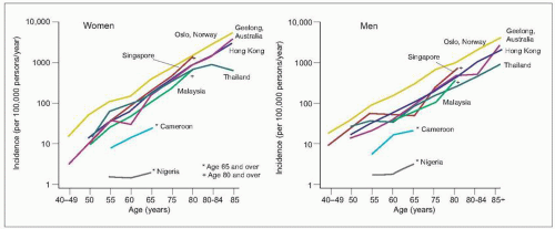
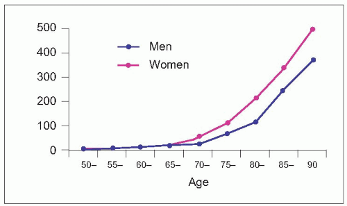
The incidence of hip fractures increases exponentially with age (2.8). Ninety percent occur in people over 50 years of age and the average age of sustaining a hip fracture in developed countries is 80 years. The estimated rates in westernized countries are 33/100,000 person-years in women aged aged 50-54 years, rising to over 1808/100,000 person-years in women 80 years and older, with rates in men of 28 and almost 900, respectively9 (Table 2.1). However, the female preponderance of hip fractures is not common to all populations. The age-specific incidence varies between countries (2.9). Worldwide, there were estimated to be 1.31 million hip fractures in 1990, about 909,000 in women and 405,000 in men10. Fracture rates vary in different countries (Table 2.1), being highest in North America and Europe, particularly in Scandinavia. The risk of osteoporotic fractures is lower in nonwhite than white populations. Incidence rates are extremely low in African countries and intermediate among Asian populations. Rates vary in regions and in Asia the rates for hip facture are twofold higher in Hong Kong than in Korea, with intermediate are reported from Malaysia, Thailand, and mainland China.
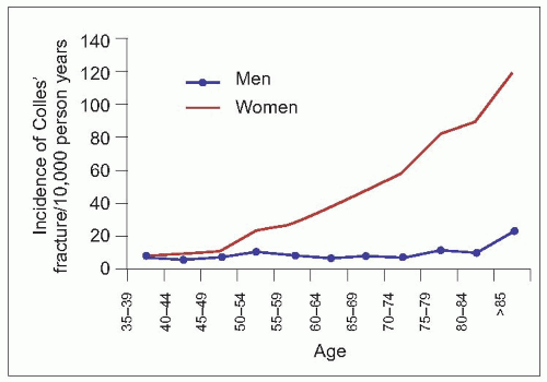
2.7 Age distribution of distal forearm fractures. (Adapted from O’Neill TW, et al. (2001). Incidence of distal forearm fracture in British men and women. Osteoporos Int 12(7):555-558.)


2.8 Age distribution of hip fractures. (Adapted from Rogmark C, et al. (1999). Incidence of hip fractures in Malmö, Sweden, 1992-1995. A trend-break. Acta Orthop Scand 70(1):19-22.)


Table 2.1 Incidence of hip fracture (rates/100,000) in 1990 by age, sex, and region


Men: age (years)


Women: age (years)


Region


50-54


55-59


60-64


65-69


70-74


75-79


80+


50-54


55-59


60-64


65-69


70-74


75-79


80+


W. Europe


28


33


67


103


203


331


880


33


54


115


184


362


657


1808


S. Europe


10


16


34


55


81


190


534


11


21


47


100


170


380


1075


E. Europe


38


38


88


88


194


194


475


58


58


155


155


426


426


1251


N. Europe


58


66


97


198


382


682


1864


74


78


190


327


612


1294


2997


N. America


33


33


81


123


119


338


1230


60


60


117


252


437


850


2296


Oceania


20


34


63


92


180


445


1157


31


63


112


204


358


899


2476


Asia


19.5


19.5


36.5


46.5


102


150


364


14


14


38


74.5


155.5


252


562.5


Africa


6


10


14


27


8


0


116


4


12


17


12


16


50


80


Latin America


25


40


40


106


106


327


327


19.5


50


50


162.5


162.5


622


622


World


22.5


24.5


47.3


68.7


119.1


219.4


630.2


23.9


28.4


69.1


121.6


239.8


457.7


1289.3


(Adapted from Gullberg B et al. (1997). Worldwide projections for hip fracture. Osteoporos Int 7:407-413.)


2.9 The age-specific incidence of hip fractures in different countries. (Adapted from Zebaze RM, Seeman E (2003). Epidemiology of hip and wrist fractures in Cameroon, Africa. Osteoporos Int 14(4):301-305.)


Other fractures

Fractures are more common with ageing at other sites, in particular the proximal humerus and pelvis. Fractures of the humerus are estimated to account for approximately 8% of all fractures sustained by adults11 and, in persons over 40 years of age, three-quarters of these are of the proximal humerus12. Data suggest that fracture of the proximal humerus is the third most common fracture over age 6511,13. Fractures of the proximal humerus have shown a similar pattern of increase with age as other common fragility fractures11 in both men and women, with women being somewhat older at the time of fracture, average around 70-74 years versus 65 years in men13. Pelvic fractures account for approximately 2% of all adult fractures11. Pelvic fractures in the elderly occur from minor trauma or from skeletal insufficiency. The majority of patients are women (up to 80%) and above the age of 80 years.


The lifetime risk of fracture is considerable. For a man or woman aged 50 years in the UK, the future lifetime risk of fracture is 20.7% and 2% respectively (Table 2.2)11. The lifetime risk of fractures from age 50 years of the forearm is 16.6% in women and 2.9% in men, whereas of the hip is 11.4% in women and 3.1% in men11. The risk of future fracture can also be expressed as a 10-year probability11 (Table 2.3), which has the advantage that it is an expression of absolute risk over a more meaningful period to when considering possible intervention. It makes it clear that the greatest potential gain from any intervention is at older ages when the absolute risk is highest. The 10-year probability will increase with increasing relative risk14 (Table 2.4). As a consequence, there is most to gain by treating older women with risk factors that increase the probability of them sustaining a fracture. Interventions then become costeffective.


Table 2.2 Estimated lifetime risks of fractures (%) in the UK at various ages


Current age (years)


Any fractures


Radius/ulna


Femur/hip


Vertebra


Women


50


53.2


16.6


11.4


3.1


60


45.5


14.0


11.6


2.9


70


36.9


10.4


12.1


2.6


80


28.6


6.9


12.3


1.9


Men


50


20.7


2.9


3.1


1.2


60


14.7


2.0


3.1


1.1


70


11.4


1.4


3.3


1.0


80


9.6


1.1


3.7


0.8


(Adapted from van Staa TP, et al. (2001). Epidemiology of fractures in England and Wales. Bone 29:517-22.)


Table 2.3 Estimated 10-year risks (%) of fractures in the UK at various ages


Current age (years)


Any fractures


Radius/ulna


Femur/hip


Vertebra


Women


50


9.8


3.2


0.3


0.3


60


13.3


4.9


1.1


0.6


70


17.0


5.6


3.4


1.3


80


21.7


5.5


8.7


1.6


Men


50


7.1


1.1


0.2


0.2


60


5.7


0.9


0.4


0.3


70


6.2


0.9


1.4


0.5


80


8.0


0.9


2.9


0.7


(Adapted from van Staa TP, et al. (2001). Epidemiology of fractures in England and Wales. Bone 29:517-22.)


Table 2.4 Probability of fracture during the next 10 years in men and women from Sweden (according to age and risk relative to the average population)


Age


Relative risk hip, clinical spine, humeral, or Colles’ fracture


50


60


70


80


(years)


10-year probability of fracture (%)


Men


1


3.3


4.7


7.0


12.6


2


6.5


9.1


13.5


23.1


3


9.6


13.3


19.4


13.9


4


12.6


17.3


24.9


39.3


Women


1


5.8


9.6


16.1


21.5


2


11.3


18.2


29.4


37.4


3


16.5


26.0


40.0


49.2


4


21.4


33.1


49.5


58.1


(Adapted from Kanis JA, et al. (2002). Ten-year risk of osteoporotic fracture and the effect of risk factors on screening strategies. Bone 30:251-58.)


Epidemiology of fracture – future trends

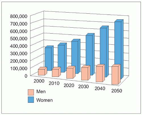
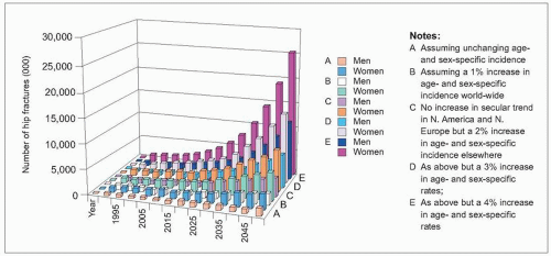
The number of hip fractures is increasing throughout the world. From an estimated 1.26 million in 1990, the projected number is 2.6 million by 2025 and 4.5 million worldwide by 20509. It is estimated that the percentage increase will be greater in men (310%) than women (240%). The increase will be in Europe (2.10) and all populations across the globe (2.11). The predicted increases in hip fractures in westernized populations is because of increased survival and growing numbers of very elderly who will be inherently at greater risk of fracture. However, it is predicted that there will be most dramatic changes in Asian populations in particular, and it has been estimated that from 26% of all hip fractures occurring in Asia in 1990, that this will rise to 37% in 2025 and to 45% in 2050. This is already being seen in the more urbanized countries of Singapore and Hong Kong where age-adjusted hip fracture rates have been found to be similar to those in white Americans, whereas much lower rates were found in Malaysia and Thailand15. Other osteoporotic fractures are also predicted to increase. This is because of the increase in populations and predicted dramatic improvements in life expectancy in less developed countries, as well as changes in lifestyles that will increase individual risk of fracture. People are becoming less physically active and changes in transportation increase the risks of trauma.


2.10 Projected number of yearly incident fractures in the European Community member states. (Adapted from European Communities (1998). Report on Osteoporosis in the European Community: action for prevention. European Communities, Luxembourg.)


2.11 Worldwide forecasts for hip fracture, 2050. (Adapted from Gullberg B, et al. (1997). Worldwide projections for hip fracture. Osteoporos Int 7(5):407-413.)


Outcome after fracture – function, mortality

The clinical manifestation of osteoporosis is fracture following low-energy trauma, such as a fall from a standing height. Fracture of a long bone will result in pain and loss of mobility of the limb. Vertebral fracture presents acutely with pain in about one-third of cases. As a consequence, function is lost, activities are limited. and participation is restricted. The person may become isolated and also fearful of falling and sustaining another fracture, of which they are at increased risk.


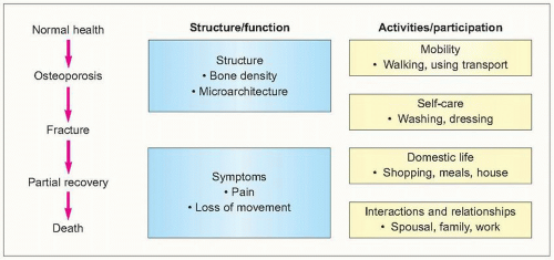
2.12 Impact of osteoporosis in terms of the WHO International Classification of Functioning.

The impact of osteoporosis and fracture can be represented in terms of the WHO International Classification of Functioning (2.12). This is a valuable model to evaluate the outcomes of osteoporosis and fracture. At the clinicopathological end of this classification, osteoporosis is characterized by loss of bone mass and architecture, and a fracture results in loss of integrity of the skeleton with pain. For the individual, a fracture not only results in pain and loss of function of that limb or structure, but will result in limitation of a wide range of activities, such as walking, and this will restrict their participation within society such as going out with friends. These physical difficulties associated with osteoporosis result in increased dependence and the enormous indirect costs of the condition. Many fractures result in hospitalization, but this will depend not only on the severity of the fracture and the impact on the individual but also on the local system of healthcare and social support.


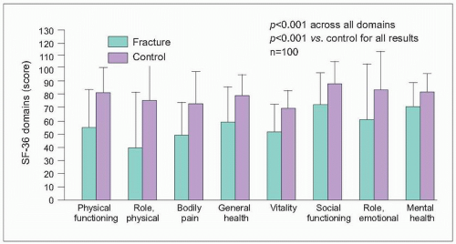
2.13 Vertebral fractures and quality of life. (Adapted from Hall SE, et al. (1999). A case-control study of quality of life and functional impairment in women with long-standing vertebral osteoporotic fracture. Osteoporos Int 9(6):508-515.)


Vertebral fractures

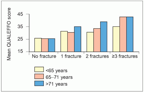
Vertebral fracture is associated with acute back pain in only one-third of fractures, but new vertebral fractures, including those that do not come to immediate clinical attention, are associated with clinically important increases in back pain and functional limitations (2.13).

Each additional new fracture is associated with further pain and limitation of activities with restriction of participation. The pain associated with an acute fracture can be severe for a couple of weeks and up to one-fifth are hospitalized and some will require subsequent long-term care. Fractures of the lower thoracic and upper lumbar spine are associated with more pain. Pain and disability worsen with each new vertebral fracture, with an increasing total number of vertebral fractures and with worsening of spinal deformity (2.14).

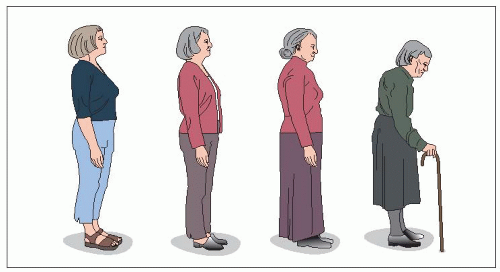
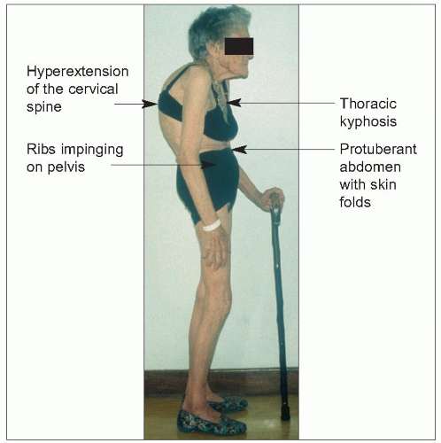
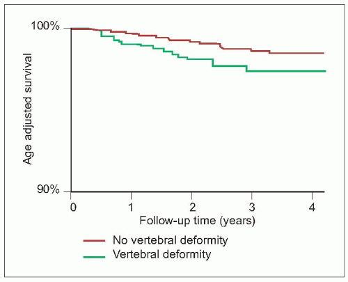
With increasing number of vertebral deformities there is loss of height and kyphosis (2.15). The abdomen becomes protuberant with increased skin folds and often intertrigo (2.16). The ribs may painfully impinge on the pelvic brim when stooping. There may be reflux oesophagitis and stress incontinence. All aspects of quality of life are affected and this is not just related to pain. Comorbidity is common at this advanced age and contributes to the impact on quality of life. Vertebral fractures are also associated with a gradual increase in mortality (2.17), in contrast to hip fractures in which the excess mortality is greatest shortly after the fracture. This increasing mortality associated with vertebral fractures may in fact be explained by confounders that relate independently to bone density and to mortality, in particular advanced age, comorbidities, and general frailty.


2.14 Impact of age and number of vertebral fractures. (Adapted from Oleksik A, et al. (2000). Health-related quality of life in postmenopausal women with low BMD with or without prevalent vertebral fractures. J Bone Mineral Res 15(7):1384-1392.)


2.15 Progressive loss of height associated with vertebral deformities.


2.16 The clinical problems associated with vertebral osteoporosis and fracture.


Distal forearm fracture

Fracture of the distal forearm can have a major effect on what the person can do (Table 2.5). In a survey in the UK, one in five men and women were admitted to hospital, more often men than women (23.4% vs. 18.6%). Below age 50 years, 22.6% of those with fracture required admission. Above 50 years the proportion admitted rose gradually with age: 14.5% at age 50-59 years, 15.9% at age 60-69 years, 17.6% at age 70-79 years, and 26% at age 80 years and over8. Many do not return to their prefracture status, with long-term limitation of function and some develop algodystrophy (reflex sympathetic dystrophy).


2.17 There is an increased mortality associated with vertebral deformities. Age-adjusted Kaplan Meier survival curves in women. (Adapted from Ismail AA, et al. (1998). Mortality associated with vertebral deformity in men and women: results from the European Prospective Osteoporosis Study (EPOS). Osteoporos Int 8(3):291-297.)


Hip fracture

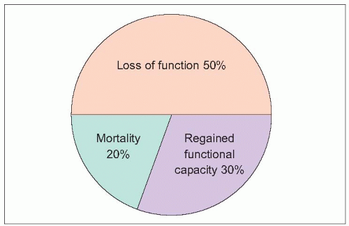
The average age of sustaining a hip fracture is 80 years in western Europe, and many are frail with comorbidities. The hip fracture results in pain, loss of mobility, and excess mortality (2.18). Nearly all are hospitalized, and most undergo surgical repair of the fracture or replacement of the joint. At 1 year, hip fracture is associated with 50% loss of function and only 30% have regained function16. Many lose their independence and require long-term care. Increasing age and comorbidity are important contributory factors to the occurrence of hip fractures and are determinants of outcome. The outcome is worse for those already in care when they sustain a fracture, but many of those who do return to their own homes do not return to their prefracture lifestyle.


2.18 Outcome after hip fracture at 1 year. (Adapted from Sernbo I, Johnell O (1993). Consequences of a hip fracture: a prospective study over 1 year. Osteoporos Int 3(3):148-153.)


Table 2.5 Distal forearm fractures (Colles)


  • 20% hospitalized


  • Require 4-6 weeks in plaster


  • Impaired function


  • Algodystrophy


One-year mortality has been found to be up to 40% in various studies. Excess mortality is greatest in the first 6 months (2.19), and in younger people17. Mortality is greater for those with coexisting illnesses and poor prefracture functional status. Major causes of death are pneumonia, pulmonary embolism, stroke, myocardial infarct, and cardiac failure.

Only gold members can continue reading. Log In or Register to continue

Stay updated, free articles. Join our Telegram channel

Jul 24, 2016 | Posted by in ORTHOPEDIC | Comments Off on Epidemiology, Risk, and Risk Factors

Full access? Get Clinical Tree

Get Clinical Tree app for offline access