CHAPTER 32 Endoscopic Release of the First Dorsal Extensor Tendon Compartment
Rationale
One might ask if arthroscopic first dorsal compartment release is a triumph of technology over reason, given that the open release is usually considered straightforward and effective. However, upon closer examination there are several reasons to consider this approach. First, although traditional open release gives generally good results complications (due to the approach) do occur and are most likely underreported. One study by Harvey et al. documented scar adherence to the underlying tendon in 2 of 20 surgical patients, 1 minor wound infection, and temporary parathesias of the radial sensory nerve in 3 patients.1 Another study by Arons et al. documents in 16 consecutive patients 14 complications, including 2 neuromas, 3 adhesions, 1 tendon subluxation, and 3 hypertrophic painful scars.2 A study by Ta et al. documented a 5% recurrence rate, 2% sensory nerve injury, and 2% with severe scar tenderness out of 43 patients.3 There have been other case reports of palmar subluxation of the tendon following operative release.4 Clearly, although an open release of the first extensor compartment is perceived as a safe, simple, and effective surgical procedure when examined closely there is room for improvement.
Surgical Technique
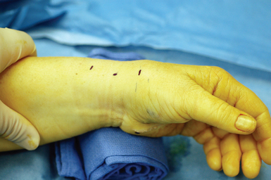
The patient is placed supine. The arm is elevated and exsanguinated, and a tourniquet inflated. The wrist is placed over a towel roll in a neutral position. A distal portal is established by making a 5-mm superficial transverse incision just distal to the thumb carpometacarpal (CMC) joint. The incision is placed in line with the first dorsal compartment at the insertion of the abductor pollicis longus (APL) tendon, 2 to 3 cm distal to the end of the radial styloid (Figure 32.1). Blunt dissection with a small hemostat is used to clear the overlying subcutaneous tissue from the fascia enveloping the thumb CMC joint, which is then elevated off the tendons of the first dorsal compartment with a small right-angle retractor.
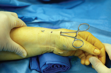
A narrow and long hemostat snap is next used to bluntly create a working space between the skin and subcutaneous tissue (Figure 32.2). A small-joint arthroscopic cannula and trocar are inserted above the fibrous fascial sheath of the first dorsal compartment, proximal to the radial styloid and the extensor tendon retinaculum. A proximal portal is established by making a second 5-mm transverse incision over the trocar tip approximately 4 to 6 cm proximal to the radial styloid (Figure 32.3). The trocar is removed and a 2.7-mm 30-degree angled scope is inserted into the cannula through the proximal portal (Figure 32.4).
< div class='tao-gold-member'>
Related posts:


Stay updated, free articles. Join our Telegram channel

Full access? Get Clinical Tree


