Fig. 9.1
(a) Changes in BMD of the lumbar spine after 5 years of treatment with alfacalcidol alone (D) or alfacalcidol and elcatonin (D + ECT). Data are presented as the mean + standard error the mean (SEM). AP anteroposterior, L lateral, Mid midlateral. *P < 0.05 and **P < 0.001 compared with the baseline BMD in each group (Reprint with permission from Springer). (b) Changes in proximal femoral BMD after 5 years of treatment with alfacalcidol alone (D) or alfacalcidol and elcatonin (D + ECT). Data are presented as the mean + SEM. FN femoral neck, TR trochanter, WD Ward’s triangle. *P < 0.001 compared with the baseline BMD in each group (Reprint with permission from Springer)
Eldecalcitol suppresses bone resorption to a greater extent than alfacalcidol and has a similar effect on bone formation and calcium metabolism, resulting in a greater increase in the BMD of OVX rats [19]. Combined therapy with eldecalcitol and alendronate improves the mechanical properties of the lumbar spine and mid-shaft femur by additive suppression of bone resorption and maintenance of bone formation in OVX rats [58, 59]. Co-treatment with eldecalcitol and raloxifene improves mechanical strength by increasing BMD in OVX rats [60]. In a clinical study, 0.75 μg eldecalcitol significantly increased lumbar and hip BMD [61]. The enhancing effects of eldecalcitol on BMD are not dependent on the baseline level of 25(OH)D [62]. A recent review of eldecalcitol showed an increase in BMD with inhibitory effects on bone resorption [63].
9.3.2 Bone Quality and Mechanical Properties
In addition to the effects of vitamin D on BMD, there have been several studies of active vitamin D effects on bone quality including mechanical properties, collagen, and collagen cross-linking. Microcomputed tomography has shown that combined therapy with etidronate and alfacalcidol increased the mechanical properties of the trabecular and cortical bone without impairment of mineralization or connectivity, resulting in bone strengthening in OVX rats [64]. In the fracture repair rat model, alfacalcidol induced lamellar bone formation coinciding with an increase in enzymatic cross-linking and normalization of the cross-linking pattern in callus to a native bone pattern [65]. Alfacalcidol also improved the amount and cross-linking pattern of collagen in steroid-treated rats [66]. A recent study has shown that alfacalcidol not only increased the amount of collagen but also enhanced the maturation of collagen in OVX rats [67]. Eldecalcitol improved the biomechanical properties of the femoral cortical bone by inhibiting endocortical bone resorption and stimulating periosteal bone formation in SAM/P6 mice [68]. In a clinical study, eldecalcitol increased the cortical cross-sectional area and maintained the cortical thickness of the proximal femur better than alfacalcidol in osteoporotic patients as evaluated by clinical computed tomography [69]. This study showed that the biomechanical properties of the femoral neck, including cross-sectional moment of inertia and the section modulus, were improved more by eldecalcitol than alfacalcidol [69]. These studies indicate that active vitamin D improves both the quality and mechanical properties of bone.
9.4 Effects on Skeletal Muscle
9.4.1 Effects of Vitamin D Deficiency on Muscle
Many studies have shown that serum levels of 25(OH)D, but not 1α,25(OH)2D3, are related to bone variables and muscle functions . Some recent studies have reported that the incidence of 25(OH)D deficiency has increased in older people [70–72]. A lower serum 25(OH)D level predicted a decrease in grip strength and appendicular muscle mass in older men and women [73]. This study indicated that the 3-year risk of sarcopenia was twofold higher in older subjects with baseline 25(OH)D levels of less than 25 nmol/L [73]. Prospective studies have also reported an association between baseline 25(OH)D levels and declines in muscle function. Wicherts et al. reported that low baseline 25(OH)D levels (<50 nmol/L) were significantly correlated with a greater 3-year decline in physical performance including a walking test, chair standing, and tandem standing in people over 65 years of age [74].
9.4.2 Effects of Vitamin D on Muscle Strength and Fatigue
In an animal study, vitamin D deficiency induced by dietary restriction and housing under incandescent lighting caused a significant reduction in muscle strength of the soleus in rats as assessed by a force transducer to detect isometric contraction [78]. We performed several animal studies to clarify the effects of alfacalcidol on muscle strength and fatigue, and the histomorphometric changes in muscle tissues of normal and OVX rats as a model for aged osteoporotic women [79, 80]. Alfacalcidol administration significantly increased the maximum contraction tension of the calf muscle in the sham-operated group (5–7 % increase, p < 0.01) and OVX group (4–7 % increase, p < 0.001) compared with their respective controls. However, alfacalcidol administration did not significantly affect muscle fatigue in these groups as evaluated by the percentage strength at each cycle of the initial contraction strength [80]. Furthermore, we evaluated the effects of alfacalcidol on skeletal muscle strength and fatigue in prednisolone-administered rats and found steroid myopathy with muscle weakness and atrophy. Alfacalcidol significantly increased the maximum contractile muscle strength (Fig. 9.2a) and decreased muscle fatigue (Fig. 9.2b) as evaluated by the strength decrement index (SDI) of the calf muscle in prednisolone-administered rats [81]. These results indicate that the active form of vitamin D, alfacalcidol, increases muscle strength and decreases muscle fatigue , resulting in prevention of falls.
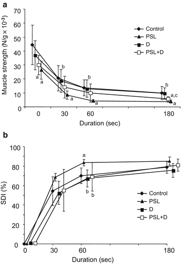
Fig. 9.2
(a) Changes in muscle strength during stimulation. Each data point for the Control (♦), PSL (▲), D (■), and PSL + D (□) groups is expressed as mean ± standard deviation (SD). a,b,c Significant differences (P < 0.05) between aControl, bPSL, and cD groups at each time point (Reprint with permission from Biomedical Research Press). (b) Changes in SDI during stimulation. Each data point for the Control (♦), PSL (▲), D (■), and PSL + D(□) groups is expressed as mean ± SD. a,b Significant differences (P < 0.05) between aControl and bPSL groups at each time point (Reprint with permission from Biomedical Research Press)
In a clinical situation, supplementation of more than 700 IU/day vitamin D led to an improvement in muscle strength and preserved bone in older people with vitamin D insufficiency (serum 25(OH)D, <50 nmol/L) [82]. Other studies also showed that vitamin D supplementation improved lower limb strength in institutionalized [83] and community-dwelling older individuals [84]. Stockton et al. reported that muscle fatigue was related to physical function but not vitamin D levels or maximal isometric strength in vitamin D-depleted patients with systemic lupus erythematosus (SLE) [85].
Treatment with 1 μg alfacalcidol increases muscle mass, muscle power, and balance and reduces fear of falling in older people [86]. Alfacalcidol (0.5 μg) has been shown to significantly improve muscle strength (isometric knee extension strength) and functional ability (walking distance over 2 min) after 6 months of treatment in older 25(OH)D-deficient women [87]. Another study demonstrated that alfacalcidol together with calcium and alendronate enhanced the beneficial effects of back extensor exercise in patients younger than those in their late 60s [88]. In addition, long-term treatment with alfacalcidol improved body sway in older women [89]. A recent study revealed that alfacalcidol maintains muscle mass and increases the skeletal muscle index in patients with low muscle mass [90].
However, a single high dose of vitamin D (300,000 IU) did not improve physical performance even in older patients with a low baseline 25(OH)D level (<12 nmol/L) [91]. A meta-analysis showed no effect on grip strength or proximal lower limb strength by vitamin D supplementation in adults with 25(OH)D levels of more than 25 nmol/L in 17 randomized controlled trials (RCTs) involving 5072 subjects [92]. Annweiler et al. also performed a systematic review to examine the effects of low serum vitamin D and vitamin D supplementation on muscles, balance, and gait performance among people aged 65 years and older [93]. They concluded that the association between vitamin D and physical performance remains controversial [93].
9.4.3 Effects of Vitamin D on Muscle Fiber Phenotypes
Vitamin D supplementation may change the muscle fiber composition. Muscle fibers are divided into type I (slow twitch) and type II (fast twitch) with further subdivision into IIA, IIX, and IIB depending on the expression of different myosin heavy chain isoforms [70]. We have recently reevaluated the effects of vitamin D on the percentages of muscle fiber phenotypes in OVX rats. After 4 weeks of oral administration of alfacalcidol (0.1 μg/kg/day) to OVX rats, their calf muscle fibers had a smaller diameter. Moreover, type I fibers increased from 83.6 to 88.3 % in OVX and control rats treated with or without alfacalcidol (Fig. 9.3a) [80]. Ovariectomy or alfacalcidol administration did not affect the smaller diameter of muscle fibers (Fig. 9.3b) [80]. Conversely, Sorenson et al. showed an increase in the relative fiber composition and size of type IIA fibers after treatment with alfacalcidol and calcium in muscle biopsies from older women [94]. A randomized controlled study found that daily treatment of older stroke patients with 1000 IU vitamin D2 increased the type II muscle fiber diameter and percentage of type II fibers over 2 years [95]. Treatment with alfacalcidol and calcium for 3–6 months increased the proportion and cross-sectional area of type IIA fibers in the vastus lateralis of aged osteoporotic patients [94].
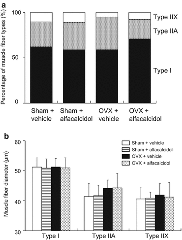
Fig. 9.3
(a) Effects of ovariectomy and/or alfacalcidol on the composition of muscle fiber types including type I (■), type IIA (▨), and type IIX (□). There was no significant difference in the composition of muscle fiber types induced by ovariectomy or alfacalcidol treatment. (b) Differences in the diameters of muscle fibers of each phenotype induced by ovariectomy (OVX) and alfacalcidol treatment. OVX or alfacalcidol administration did not affect the diameter of muscle fibers in any of the four groups
9.4.4 Effects of Eldecalcitol on Muscle
Eldecalcitol increases the expression of several factors related to muscle function in C2C12 cells in vitro, such as MyoD and myogenin [96]. To evaluate in vivo effects of eldecalcitol on skeletal muscle morphology and function, we treated glucocorticoid-induced myopathic and osteopenic rats with eldecalcitol. Four weeks of treatment with eldecalcitol prevented muscle atrophy of the tibialis anterior and loss of femoral BMD and increased calf muscle strength. However, there was no significant preventive effect on muscle fatigue induced by eldecalcitol [97]. A recent study has demonstrated that eldecalcitol enhances the expression of insulin-like growth factor-1 (IGF-1), myelin basic protein, and VDR in rat primary Schwann cells, and VDR signaling induced by eldecalcitol regulates neuromuscular maintenance and enhances locomotive ability following physical exercise [98].
In a clinical situation, eldecalcitol improved muscle power as evaluated by chair-rising time in postmenopausal women with osteoporosis treated with alendronate or risedronate [99]. We have also performed a prospective study to evaluate the effects of eldecalcitol on static and dynamic body balance in older osteoporotic women. Eldecalcitol and alendronate co-treatment improved muscle strength measured at the back extensor and dynamic body balance as evaluated by dynamic sitting balance and a timed up and go test [100]. These results indicate that eldecalcitol also exerts significant effects on muscle strength, body balance, and physical functions .
9.4.5 Genomic and Non-Genomic Effects on Muscle
As a genomic effect on muscle, it has been reported that 1α,25(OH)2D3 regulates muscle calcium uptake [101], affects the synthesis of muscle cytoskeletal proteins [102], and regulates phosphate metabolism in myoblasts [102]. Recently, it was found that 1α,25(OH)2D3 also affects muscle function through a transcription-enhancing effect on proteins such as IGF-1 and its binding proteins as well as proteins involved directly in calcium metabolism, revealing an anabolic effect on muscle tissue [103]. Eldecalcitol induces expression of MyoD and myogenin through induction of osteoglycin, which is secreted from myoblast and stimulates osteoblastic differentiation, in C2C12 cells when combined with 1α,25(OH)2D3 [96]. However, as a non-genomic effect on muscle, 1α,25(OH)2D3 activates protein kinase C (PKC) to release calcium into the cytosol [104]. These studies suggest that alfacalcidol also has positive effects on muscle strength via genomic and non-genomic effects on muscle functions.
9.5 Effects on Falling
9.5.1 Vitamin D Deficiency and Falls
Approximately 30 % of community-dwelling people over the age of 65 years fall each year [105]. Falls are a major risk factor for fracture and other injuries and worsen quality of life [106]. Several studies have demonstrated that low baseline 25(OH)D levels are increased in older people at risk of subsequent falls [107, 108]. Older people in nursing homes and hostel residents who fall have lower serum 25(OH)D levels and higher serum PTH levels than other residents [109]. These studies indicate that low serum 25(OH)D levels are related to falling, especially in older people.
9.5.2 Prevention of Falls by Vitamin D Treatment
Although native vitamin D exerts small effects on BMD, many studies have demonstrated a beneficial effect of native vitamin D on fractures by prevention of falls [110–112]. In 2004, Bischoff et al. performed a meta-analysis based on five RCTs including people of more than 60 years of age (total number of subjects, 1237) to evaluate the effects of native vitamin D on falls [110]. They found that native vitamin D significantly reduced falls by 22 % (95 % CI, 0.64–0.92). The study also reported that treatment of 15 subjects with native vitamin D would prevent one fall [110]. Another meta-analysis with eight RCTs also demonstrated that native vitamin D treatment at more than 700 IU/day significantly reduced falls with a relative risk (RR) of 0.81 and 95 % CI of 0.77–0.92 [2]. The most recent meta-analysis included 26 RCTs with 45,782 participants, revealing that native vitamin D significantly reduced the risk of falls [odds ratio (OR) for suffering at least one fall, 0.86; 95 % CI, 0.77–0.96] [112].
Conversely, there have been several negative randomized studies between vitamin D supplementation and falls. Vitamin D (400 IU/day) treatment had no significant effect on falls among 354 older persons in the Netherlands [113]. Although a high dose of vitamin D (800 IU/day) showed positive effects on the risk of falls in nursing home residents, lower doses of vitamin D (200, 400, and 600 IU/day) did not demonstrate significant effects on the risk of falls [114]. Another two studies with larger numbers of subjects did not find a beneficial effect of 800 IU/day vitamin D on falls in people over 70 years of age or in older women living in nursing homes [115, 116]. However, a subgroup analysis of a recent meta-analysis demonstrated that the effect of vitamin D on prevention of falls was more significant in subjects with vitamin D deficiency (OR, 0.53; 95 % CI, 0.39–0.72) than in those with sufficient vitamin D (OR, 0.90; 95 % CI, 0.81–0.99). A high dose (>800 IU/day) of vitamin D (OR, 0.82; 95 % CI, 0.73–0.93) was more effective than a lower dose (<800 IU/day) of vitamin D (OR, 1.00; 95 % CI, 0.72–1.37). Furthermore, combined therapy with vitamin D and calcium (OR, 0.83; 95 % CI, 0.72–0.93) was more beneficial than monotherapy with vitamin D (OR, 0.97; 95 % CI, 0.84–1.11) [112].
Active vitamin D3 also inhibits osteoporotic fractures by acting on bone tissue and improves muscle strength and sense of balance, thus helping to prevent falls [2, 117]. Alfacalcidol has been shown to decrease postural sway in older people during 12 months of treatment [118]; improve muscle power and balance, as evaluated by a timed up and go test; and reduce the incidence of falls [119]. Studies have also demonstrated the positive effects of eldecalcitol on physical functions [99] and body balance [100]. These effects of eldecalcitol on muscle and physical functions might contribute to reducing the risk of falls. In 1054 osteoporotic patients, the incidence of wrist fractures caused by falls was significantly lower in the eldecalcitol-treated group compared with that in the alfacalcidol-treated group after 36 months [120].
Based on these results, vitamin D supplementation and active vitamin D, including eldecalcitol, are probably effective in conjunction with calcium to prevent falls among older individuals. The effects appear to be optimal in those who are vitamin D deficient at baseline.
9.6 Preventive Effects on Osteoporotic Fractures
9.6.1 Vitamin D Deficiency and Fractures
Serum 25(OH)D level is related to the muscle strength [73, 74] and the incidence of falls [107–110]. Furthermore, serum 25(OH)D levels influence the risk of osteoporotic fractures. Serum 25(OH)D concentrations of 40 nmol/L or less result in a marked increase in the risk of hip fracture s in older people, which is unrelated to their serum 1α,25(OH)2D3 levels [121, 122]. Meta-analyses of case-control studies have clearly demonstrated that a decrease in the serum 25(OH)D level is significantly associated with an increased risk of hip fractures among older people [123]. Conversely, this relationship is not obvious between the serum 1α,25(OH)2D3 level and hip fractures [124].
9.6.2 Prevention of Fractures by Vitamin D Treatment
The most important goal of osteoporosis treatment is prevention of fractures including vertebral and non-vertebral fractures in older people. Vitamin D has obvious positive effects on both bone and skeletal muscle as shown by previous studies. There have been several RCTs that evaluated the effects of vitamin D supplementation and active vitamin D on prevention of osteoporotic fractures.
Papadimitropoulos et al. performed a meta-analysis of the preventive effects of vitamin D supplementation and active vitamin D on osteoporotic vertebral fractures . Their study demonstrated that vitamin D significantly reduced the risk of vertebral fractures (RR, 0.63; 95 % CI, 0.45–0.88) [1]. In terms of the preventive effects of vitamin D supplementation on non-vertebral fractures, Bischoff-Ferrari et al. reported a meta-analysis including 12 double-blind RCTs. Their study showed that vitamin D supplementation significantly decreased the risk of non-vertebral fractures (RR, 0.86; 95 % CI, 0.77–0.96) and hip fractures (RR, 0.91; 95 % CI, 0.78–1.05) [111]. A recent analysis including 11 double-blind RCTs of 31,022 people (65 years of age or older) with 1111 incident hip fractures and 3770 non-vertebral fractures demonstrated that the highest doses (800–2000 IU/day) of vitamin D supplements reduced the risk of hip fractures by 30 % and any non-vertebral fracture by 14 % in older people [125] (Table 9.1). Another study showed that supplementation with both vitamin D and calcium improved hip BMD but had no effect on the risk of fracture [126]. It has been speculated that the effects of vitamin D supplementation on the risk of osteoporotic fractures may depend on the background of the subjects, such as age, baseline serum 25(OH)D level, and past history of osteoporotic fractures. A recent meta-analysis also reported no significant interactions between the highest actual intake of vitamin D and subgroups defined by age, type of dwelling, baseline 25(OH)D level, and additional calcium intake [125]. Instead of 25(OH)D, 1α,25(OH)2D3 administration showed no reduction in fracture risk in a meta-analysis [127].
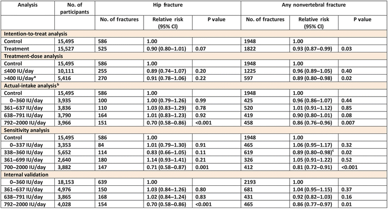
Table 9.1
Fracture incidence among 31,022 participants, according to the vitamin D treatment dose and actual intake
Similar to native vitamin D, active vitamin D, calcitriol, also significantly reduced the incidence of osteoporotic vertebral fractures in older people [128, 129]. A 2-year double-blind study by Shiraki M et al. demonstrated that alfacalcidol decreased new osteoporotic fractures in one third of the control group [53]. A meta-analysis demonstrated that active vitamin D reduced the risk of vertebral fractures with an RR of 0.53 and non-vertebral fractures with an RR of 0.34 [130]. Furthermore, the effects of active vitamin D on the reduction of fracture risks are better than native vitamin D [131].
A new analog of the active form of vitamin D, eldecalcitol, has been approved for the treatment of osteoporosis in Japan since 2011. A phase III clinical trial of eldecalcitol for osteoporosis showed a significant reduction in the incidence of new vertebral fractures over 3 years (hazard ratio (HR), 0.74; 90 % CI, 0.56–0.97) compared with alfacalcidol [120]. The incidence of new severe vertebral fractures (grade III by semiquantitative classification) in the eldecalcitol treatment group was significantly lower than that in the alfacalcidol treatment group (HR, 0.53; 95 % CI, 0.29–0.96) [132]. Compared with alfacalcidol, the risk of non-vertebral osteoporotic fractures was significantly reduced by eldecalcitol (HR, 0.59; 95 % CI, 0.37–0.94) [133]. It has been also reported that eldecalcitol significantly reduces the risk of forearm fractures compared with alfacalcidol [120].
Based on these results, vitamin D supplementation and active vitamin D have significant preventive effects on osteoporotic vertebral and non-vertebral fractures.
9.7 Conclusion
Vitamin D increases BMD and mechanical properties and improves muscle strength and physical functions. Native and active forms of vitamin D are useful to prevent falls as well as osteoporotic vertebral and non-vertebral fractures by acting on both bone and skeletal muscle in older people. A new analog of the active form of vitamin D, eldecalcitol, also increases BMD and improves physical functions. Eldecalcitol reduces the incidence of vertebral and wrist fractures more than the conventional active form of vitamin D. These effects of vitamin D result in a decrease in falls and osteoporotic fractures.
References
1.
Papadimitropoulos E, Well G, Shea B et al (2002) Osteoporosis Methodology Group and The Osteoporosis Research Advisory Group. Meta-analyses of therapies for postmenopausal osteoporosis. VIII: meta-analysis of the efficacy of vitamin D treatment in preventing osteoporosis in postmenopausal women. Endocr Rev 23:560–569PubMed
2.
Bischoff-Ferrari HA, Dawson-Hughes B, Staehelin HB et al (2009) Fall prevention with supplement and active forms of vitamin D: a meta-analysis of randomized controlled trials. Br Med J 339:b3692. doi:10.1136/bmj.b3692
4.
DeLuca HF (2004) Overview of general physiologic features and functions of vitamin D. Am J Clin Nutr 80:1689S–1696SPubMed
Related posts:
 Vertebral Strength Changes as Assessed by Finite Element Analysis
 Spondylosis and Osteoporotic Vertebral Fractures
 Applications of Teriparatide for Fracture Repair and Osteosynthetic Surgery in Osteoporosis
 Collagen Cross-Links as a Determinant of Bone Quality
 Sarcopenia and Osteoporosis
 Back Extensor Strengthening Exercise and Osteoporosis
Stay updated, free articles. Join our Telegram channel

Full access? Get Clinical Tree


