Fig. 16.1
Medium palmar aponeurosis
It spreads between the thenar and hypothenar aponeurosis, following the palmaris longus.
It’s divided in four pre-tendinous strips that change direction at the level of the distal palmar crease to form various fibrous planes:
Superficial, attaching to the dermis between the distal palmar crease and the proximal digital flexion crease (the crease of the metacarpophalangeal flexion)
Medium that extends along each of the sides of the metacarpophalangeal joints to participate in the formation of the digital aponeurosis of the corresponding finger
Deep, attaching to the palmar side of the metacarpophalangeal joint and more distally on the flexor sheath (at the level of the C1 pulley)
There are also some vertical partitions at the level of the proximal palmar crease, the partitions of Legueu and Juvara, connecting the medium and deep palmar aponeurosis.
These partitions delimit seven areas that contain alternatively the flexor system and the neurovascular pedicles with the corresponding lumbrical (Fig. 16.2).
Fig. 16.2
Partitions of Legueu and Juvara delimiting alternatively the flexor system and the neurovascular pedicles + lumbrical
16.2.2 Digital Aponeurosis [6]
The medium place of the pre-tendinous strips forms two bundles laterally prolonging toward the corresponding finger to form the sagittal strips of the finger.
These sagittal strips receive expansions from the natatory ligament to form the digital blade that protects the digital neurovascular system and fixes the cutaneous coating (Fig. 16.3).
Fig. 16.3
At the level of the distal palmar crease, the medium fibrous plane of the palmar aponeurosis joins the expansions of the natatory ligament to form the digital blade
This blade emits fibers at the level of the proximal interphalangeal joint to form the retrovascular strip, the Cleland ligament, and the Grayson ligament.
At the level of the 2nd phalanx, its anterior fibers form the oblique retinacular ligament.
We should, however, individualize some of the elements of this aponeurosis:
The oblique retinacular ligament, anchored on the flexor sheath next to the 1st phalanx diaphysis. It passes in front of the flexion axis of the proximal interphalangeal joint and joins the extensor system at the level of the distal interphalangeal joint. This ligament allows the coordination between the distal and proximal interphalangeal joints in flexion and extension. During the flexion of the distal interphalangeal joint, the oblique retinacular ligament gets tensed, automatically causing the flexion of the proximal interphalangeal joint. During the extension of the proximal interphalangeal joint, the oblique retinacular ligament gets tensed, automatically causing the extension of the distal interphalangeal joint (Fig. 16.4).
Fig. 16.4
The oblique retinacular ligament favors the functional coordination between the distal and proximal interphalangeal joints. The extension of the proximal interphalangeal joint puts it in tension, which leads to the extension of the distal interphalangeal joint. Meanwhile, the flexion of the distal interphalangeal joint is only possible if the proximal interphalangeal joint is flexed (relaxation of the oblique retinacular ligament)
The transverse retinacular ligament: its proximal half is anchored on the A3 pulley, and its distal half on the volar plate and the capsule of the proximal interphalangeal joint. It goes vertically toward the lateral strip of the corresponding extensor system. It favors the palmar movement of the lateral strips during the flexion of the proximal interphalangeal joint (Fig. 16.5). It forms the lateral retinacular ligament (with the oblique retinacular ligament).
Fig. 16.5
The transverse retinacular ligament participates in the stabilization of the lateral strips of the extensor system, opposing their dorsalization (up). The triangular ligament opposes the ventralization of the lateral strips (down)
The dorsal retinacular ligament: composed by the median strip and the arch-shaped fibers, it’s tensed between the two strips of the extensor system. It favors the dorsal movement of the lateral strips during the extension of the proximal interphalangeal joint and limits their palmar gliding during the flexion (Fig. 16.5).
The Cleland ligament: laterally tensed between the osseous skeleton and the dermis, it creates a frontal partition behind the neurovascular pedicle. In addition to its role of protecting the neurovascular pedicle, it fixes the dermis to the fibrous skeleton. Some authors don’t differentiate it from the retrovascular strip (Fig. 16.6).
Fig. 16.6
Cleland and Grayson ligaments. (AV) forward, (AR) backward
The retrovascular strip: from the digital blade at the level of the proximal interphalangeal joint, it partitions the neurovascular pedicle (Fig. 16.6).
The Grayson ligament: from the digital blade at the level of the proximal interphalangeal joint, it partitions the neurovascular pedicle (Fig. 16.6).
The longitudinal fibers: next to the flexion creases of the fingers (between the fat pads), they fix the dermis to the fibrous skeleton, allowing an efficient grip.
16.2.3 Aponeurosis System of the 1st Finger (Fig. 16.7)
Fig. 16.7
Aponeurosis system of the 1st finger (According to M. Merle)
The digital structures of the thumb can be compared to the ones of the long fingers, but we can still observe some differences.
The radial fibers of the palmar aponeurosis go toward the thumb and split into two planes (three in the long fingers), one superficial for the dermis and the other deeper for the flexor pollicis longus sheath.
The natatory ligament forms, on the radial side, the distal commissural ligament that inserts on each side of the flexor pollicis longus sheath.
The superficial transverse ligament forms, on the radial side, the proximal commissural ligament that inserts on the flexor pollicis longus sheath. With the end of the distal commissural ligament, it forms an important fibrous knot on the base of the thumb.
16.3 Anatomopathology
We describe three stages:
Initial stages: fibroblastic proliferation
Intermediate stage: fibroblastic and organization of the collagen by myofibroblastic proliferation
Terminal stage: organization of mature collagen
16.3.1 Anatomical Expression [7]
The nodules: essentially palmar; they progressively invade the tissues and strongly stick to the skin.
They are never responsible for articular retraction.
The cords: they lead to articular retractions and are the expression of this pathology.
We can find pre-tendinous palmar cords and digital cords (median, spiral, lateral, and retrovascular).
They cause the retraction of the metacarpophalangeal, the proximal interphalangeal, and rarely the distal interphalangeal (which can even be in hyperextension because of a boutonnière deformity).
The proximal interphalangeal tends to get stiff, opposite to the metacarpophalangeal.
Anatomical repartition: by order of decreasing frequency for the long fingers (V, IV, III, II).
The thumb is damaged in 10–35 % of the cases.
16.3.2 Epidemiology [8]
Dupuytren disease is more frequent in white men from Northern Europe [9].
It is unusual in black people. In 25 % of the cases, there is a hereditary factor, related to a dominant autosomal recessive gene.
16.3.2.1 Related Affections [10]
Alcoholism doubles the risks, which are even more important in case of alcoholic cirrhosis.
Diabetes, Dupuytren is related to the duration of diabetes. After 20 years of evolution, 67 % of the diabetic patients have a Dupuytren disease. However, it is often limited and almost never needs surgery.
Epilepsy doubles the risks.
Traumas and manual activities: there are no statistic proofs, but it could be a triggering factor in patients with a predisposition.
16.3.2.2 Diathesis [11]
The pathology will be more severe and recurring if it starts early and is bilateral and if there is a hereditary and/or ethnic characteristic.
The association with knuckle pads, plantar fibromatosis, and damage in the lateral fingers are pejorative factors.
16.4 Clinical and Paraclinical Signs [12, 13]
The diagnostic can be done with a clinical exam alone.
We systematically observe retractions in flexion of the metacarpophalangeal and proximal interphalangeal that can be associated with a hyperextension of the distal interphalangeal.
The palpation shows a thickening of the damaged tissues and indurations at the level of the cords and nodules.
The natatory ligament is damaged (or distal commissural ligament in the thumb), so the commissural opening decreases, which can cause maceration.
If the patient can’t put his hand flat on the table (Fig. 16.8), we should take a specialized advice to avoid important tissue retractions that can make the surgery complicated.
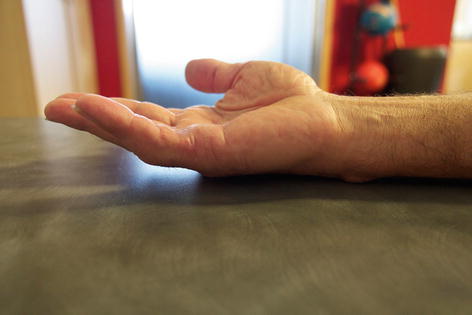
Fig. 16.8
Test of holding the hand flat on the table
The topography of the injuries, which depends on the damaged elements of the aponeurosis, is resumed in the grading system of Tubiana and Michon (Fig. 16.9).
Fig. 16.9
Grading system of Tubiana and Michon
Some rare manifestations of this pathology are:
Knuckle pads or dorsal nodules located next to the proximal interphalangeal joint that adhere to the skin but not to the extensor system. They can be painful and functionally disturbing.
Peyronie disease, retraction of a conjunctive tissue at the level of the albuginea that creates a dorsal angulation in erection.
Plantar fibromatosis, plantar equivalent of the Dupuytren and Peyronie disease.Related posts:

Stay updated, free articles. Join our Telegram channel

Full access? Get Clinical Tree


