24 Distal Ulna Implant Arthroplasty
Indications
- Chronic instability of the distal ulna after Darrach resection (see Chapter 21)
- Osteoarthritis or posttraumatic arthritis in patients with low activity demands
Pitfall
Implant stability may be difficult to achieve in cases with preoperative radioulnar divergence.
Technique
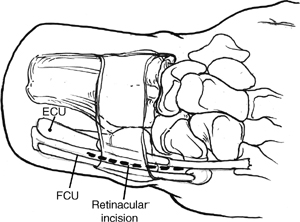
- The skin is incised along the subcutaneous border of the ulna, extending 6 to 8 cm proximally from the triquetrum (Fig. 24-1).
- Identify and protect the dorsal sensory branch of the ulnar nerve.
- The extensor retinaculum is incised between the extensor carpi ulnaris (ECU) and flexor carpi ulnaris (FCU).
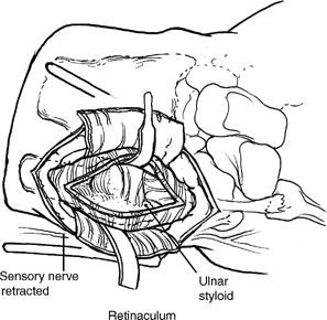
- Elevate the ECU sheath subperiosteally from the distal ulna along with the triangular fibrocartilage complex (TFCC) and other soft tissues (Fig. 24-2).

Figure 24-1
Pearl
A dorsal approach is particularly useful when there is a preexisting dorsal incision or if joint inspection is required to decide optimal treatment.
- Use radiographs and templates to choose the proper head size.
- A resection guide is used to determine the osteotomy level through the ulnar neck.
- The distal ulna is removed, preserving all soft tissues, which will form a pocket for the prosthetic head (Fig. 24-3).


Stay updated, free articles. Join our Telegram channel

Full access? Get Clinical Tree