Distal Hamstring Lengthening
Jon R. Davids
DEFINITION
The gait pattern of children with cerebral palsy (CP) is frequently disrupted by dynamic overactivity and shortening of the medial hamstring muscles.
This disruption is characterized by increased knee flexion in stance phase and decreased knee extension at the end of swing phase.
Surgical lengthening of the medial hamstrings is usually performed in conjunction with other surgical procedures selected to address all elements of soft tissue and skeletal dysfunction that compromise gait in children with CP.
This surgical strategy, termed single-event multilevel surgery (SEMLS), requires a comprehensive assessment of gait dysfunction using quantitative gait analysis.
Proper management (surgical, orthotic, and rehabilitative) in childhood can result in an improved gait pattern that will be sustainable in the adult years.
ANATOMY
The medial hamstrings consist of three muscles: the gracilis, the semimembranosus, and the semitendinosus. All three are considered biarticular muscles because they cross both the hip joint and knee joint.
The gracilis muscle is innervated by the obturator nerve and has its origin on the inferior pubic ramus and its insertion on the anteromedial aspect of the proximal tibia. It serves as a hip adductor and knee flexor. The gracilis muscle has a relatively small physiologic cross-sectional area and a relatively large ratio of tendon length to muscle fiber length, indicating that it is designed for maximal excursion and diminished force generation.8, 11
The semimembranosus muscle is innervated by the sciatic nerve and has its origin on the inferolateral portion of the ischium and its insertion on the posteromedial aspect of the proximal tibia. It serves as a hip extensor and knee flexor. The semimembranosus muscle has a relatively large physiologic cross-sectional area and a relatively small ratio of tendon length to muscle fiber length, indicating that it is designed for minimal excursion and increased force generation.8, 11
The semitendinosus muscle is innervated by the sciatic nerve and has its origin on the inferomedial portion of the ischium and its insertion on the anteromedial aspect of the proximal tibia. It serves as a hip extensor and knee flexor. The semimembranosus muscle has a relatively small physiologic cross-sectional area and a relatively large ratio of tendon length to muscle fiber length, indicating that it is designed for maximal excursion and diminished force generation.8, 11
The lateral hamstrings consist of the biceps femoris muscle, which is innervated by the sciatic nerve. The muscle is considered to be uniarticular, crossing only the knee joint, with its origin on the posterior aspect of the middle third of the femur and its insertion on the fibular head. It serves as a knee flexor. The biceps femoris muscle has a relatively large physiologic cross-sectional area and a relatively small ratio of tendon length to muscle fiber length, indicating that it is designed for minimal excursion and increased force generation.8, 11
PATHOGENESIS
CP is the consequence of an injury to the immature brain that may occur before, during, or shortly after birth. The nature and location of the injury to the central nervous system (CNS) determines the neuromuscular and cognitive impairments.
Common functional deficits are related to spasticity, impaired motor control, and disrupted balance and body position senses.
Although the injury to the CNS is not progressive, the clinical manifestations of CP change over time because of growth and development of the musculoskeletal system. The muscles typically exhibit a purely dynamic dysfunction during the first 6 years of life, characterized by a normal resting length and an exaggerated response to an applied load or stretch. With time, between 6 and 10 years of age, the muscles develop a fixed or myostatic shortening, resulting in a permanent contracture.
As such, it is best to consider CP not as a single specific disease process but rather a clinical condition with multiple possible causes.16
NATURAL HISTORY
Ambulatory children with CP whose gait is disrupted by overactivity and shortening of the medial hamstring muscles typically walk with increased knee flexion in stance phase and diminished knee extension in swing phase.
This is usually associated with increased ankle plantarflexion in stance phase and is called a jump gait pattern.14, 18
Children with a jump gait pattern will have overactivity of the medial hamstring muscle group; the lateral hamstring muscle group is rarely involved.
As the child gets older and heavier, ankle plantarflexor insufficiency (due to muscle weakness and foot segmental malalignment) will eventually occur. This will result in increasing ankle dorsiflexion and knee flexion in stance phase, which is called a crouch gait pattern.14, 18
Teenagers with a severe crouch gait pattern will have involvement of both the medial and lateral hamstring muscle groups.
As the ground reaction force falls further and further behind the knee during the stance phase in crouch gait, the demands
on the knee extensor muscles increase, eventually resulting in painful patellofemoral overload.
For this reason, the crouch gait pattern is not sustainable, and by the late teenage or young adult years, individuals with this gait pattern frequently lose the ability to ambulate.
PATIENT HISTORY AND PHYSICAL FINDINGS
The clinical history, as provided by the child and the parents, usually contains complaints of inability to stand up straight, inability to keep up with peers in play and sports, inability to walk distances (such as at the grocery store or the mall), and anterior knee pain after walking activities or at the end of the day.
A straight-leg raise of 60 degrees or less is indicative of shortening of the medial hamstrings.
A popliteal angle of 45 degrees or greater is indicative of shortening of the medial hamstrings.
IMAGING AND OTHER DIAGNOSTIC STUDIES
Radiographic imaging is not required when determining the need for lengthening of the medial hamstrings to improve gait in children with CP.
If lateral radiographs of the knee are obtained, however, they will frequently show patella alta, with fragmentation of the inferior pole of the patella; these are the sequelae of chronic and progressive patellofemoral overload (FIG 1).
Relevant data from quantitative gait analysis include sagittal plane kinematics and kinetics at the knee and dynamic electromyography (EMG) of the medial hamstrings.
Sagittal plane knee kinematics will show increased knee flexion of greater than 20 degrees at initial contact in loading response at the beginning of stance phase and diminished knee extension at terminal swing at the end of swing phase (FIG 2A).5 Midstance alignment of the knee may be variable.
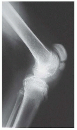
FIG 1 • Standing lateral radiograph of the knee in a child with CP and a crouch gait pattern. There is a patella alta, with fragmentation of the inferior pole of the patella, indicating chronic overload of the knee extensor mechanism.
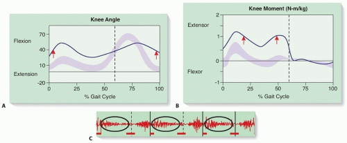
FIG 2 • Sagittal plane knee kinematic plots of a child with a jump gait pattern (A) and kinetic plots of a child with crouch gait pattern (B). The age-matched normal motion (mean ± 2 standard deviations) appears as a light purple band, and the subject’s data are indicated by a blue line. A. The gait cycle is on the horizontal axis, the direction of motion on the vertical axis. Kinematic indicators for lengthening of the medial hamstrings are increased flexion at initial contact and diminished extension at terminal swing (arrows). B. The gait cycle is on the horizontal axis, the internal moment on the vertical axis. Crouch gait pattern is characterized by an increased internal extension moment at the knee throughout stance phase (red arrows). C. Dynamic EMG of the medial hamstrings in a child with CP. Three gait cycles are shown, separated by the solid vertical lines. The stance and swing phases of each cycle are separated by the dashed vertical lines. The normal timing of activation of the muscle is noted by the horizontal red lines at the bottom of the strip. The actual timing of activation of the muscle for the subject is shown by the oscillating red line at the middle of the strip. The dynamic EMG indicator for lengthening of the medial hamstring muscles is prolonged activity in midstance (indicated by the circles in each gait cycle).
In jump gait pattern, full knee extension in midstance may occur. This is not a contraindication to lengthening of the medial hamstrings.
In crouch gait pattern, increased knee flexion will be of greater magnitude and is present throughout the stance phase.
Sagittal plane knee kinetics will show an increased internal knee extension moment in stance phase (FIG 2B).12
In jump gait pattern, the increased knee moment will occur in the loading response and terminal stance subphases of stance phase.
In crouch gait pattern, the increased knee moment will be of greater magnitude and will occur throughout the stance phase.
Dynamic EMG of the medial hamstrings will show prolonged activity of the muscle group into the mid- and terminal stance subphases of stance phase (FIG 2C).
DIFFERENTIAL DIAGNOSIS
Increased knee flexion in stance phase may be the consequence of the following:
Overactivity or shortening of the medial hamstring muscles
Surgical lengthening of the medial hamstring muscles is appropriate.
Ankle plantarflexor insufficiency, with disruption of the ankle plantarflexion-knee extension couple. This may be a consequence of muscle weakness or foot skeletal segmental malalignment, resulting in lever arm deficiency.7, 10, 17
The ankle plantarflexor insufficiency must be addressed directly to improve the gait deviation at the knee.
Dyskinetic CP, with disrupted balance and body position sense. In such cases, mildly increased knee flexion in stance phase gives a sense of stability to the child and is habitual.
Surgical lengthening of the medial hamstring muscles will not correct the gait deviation in this situation.
Increased knee flexion at the end of swing phase may be a consequence of the following:Related posts:

Stay updated, free articles. Join our Telegram channel

Full access? Get Clinical Tree


