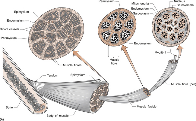
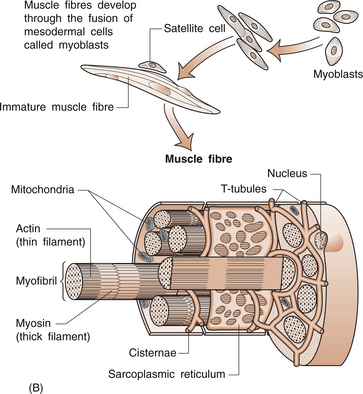
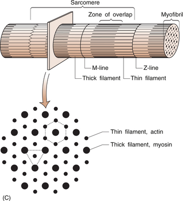
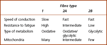
DISORDERS OF SKELETAL MUSCLE

8


DISORDERS OF SKELETAL MUSCLE


Rodger Laurent


Introduction


Skeletal muscle is one of the major tissue components of the human body. Its main function is to convert chemical energy into mechanical work. It is usually under the voluntary control of the central nervous system.


Skeletal muscle also has other functions; it contains about 80% of the body’s content of water and is a reservoir for intracellular ions such as potassium. It also functions as a source of energy-rich compounds and is an important producer of body heat.


A myopathy is a primary disorder of the muscle. The acquired myopathies include polymyositis, and endocrine and drug-induced myopathies. The inherited myopathies are rare but need to be considered in someone presenting with weakness because they often have a poor prognosis.


This chapter will discuss the primary muscle disorders. It will not discuss muscle disorders secondary to nerve or motor endplate disorders.


Anatomy of skeletal muscle


The muscle fibre


Muscles are made up of a collection of individual muscle fibres (Figs 8.1, 8.2). Each fibre is a multinucleated cell, which can be up to 10 cm in length with a diameter ranging from 10–100 μm. Normal muscles have the nuclei arranged around the periphery of the cells.


The muscle cell membrane is called the sarcolemma and the cytoplasm, the sarcoplasm. The sarcolemma has the property of excitability and can conduct the electrical impulses that occur during depolarization. A system of tubules, the transverse tubules or T-tubules, begins at the sarcolemma and extends into the sarcoplasm. They allow rapid distribution of the signal to contract throughout the muscle fibre.


The muscle fibre contains numerous myofibrils, which are 1–2 μm in diameter and the length of the cell (Fig. 8.1A). Myofibrils shorten and are the structures responsible for muscle contraction. They shorten the fibre because they are attached to the sarcolemma at each of its ends. Mitochondria and glycogen granules are situated between the myofibrils.


The myofibrils consist of bundles of filaments, which are made up of the proteins actin and myosin, and are organized in repeating functional units called sarco-meres. Sarcomeres are the smallest functional units of the muscle fibre (Fig. 8.1C). The actin filaments are thin and the myosin filaments are thick. The thick filaments lie at the centre of the sarcomere with the thin filaments at either end. On either side of the centre, there is an area of overlap between the thin and thick filaments in which each myosin filament is surrounded by a hexagonal array of actin filaments. The arrangement of the myosin and actin filaments gives a banded appearance to the muscle and is the reason it is called striated muscle. The sarco-meres are separated by a dense area called the Z line. The M line is at the middle of the sarcomere and consists of proteins that bind the thick myosin filaments. The actin and myosin filaments are joined by molecular cross-bridges and, during contraction, these cross-bridges repeatedly disengage and engage at successive sites, with the result that the actin and myosin filaments slide upon one another and the myofibrils shorten.


The myofibril is surrounded by a sheath of membranes called the sarcoplasmic reticulum. At the zone of overlap between the thick and thin filaments, the tubules of the sarcoplasmic reticulum enlarge and form chambers called terminal cisternae. A transverse tubule is situated between two terminal cisternae and the resulting complex is called a triad. The cisternae contain large stores of calcium ions. Release of calcium from these structures initiates the muscle contraction.


The size of the muscle varies in proportion to the size of the fibres with larger fibres being present in larger muscles. Consistent physical exercise can increase the muscle fibre diameter in both sexes.


Skeletal muscle structure


Each fibre is surrounded by a thin layer of collagen, called the endomysium. The fibres are then joined together in bundles to form fascicles, which are surrounded by a further layer of connective tissue called the perimysium (Fig. 8.1A). Groups of fascicles form the whole muscle, which is surrounded by a strong layer of collagen, called the epimysium. The epimysium merges with the peritenon of the tendon and the periosteum.


The arrangement of the fascicles is variable and depends on the task of that specific muscle. Factors such as the amount and direction of the force required or the amount of muscle shortening determine the muscle architecture. Two examples are an arrangement of the fascicles parallel to the long axis of the muscle, as in the gastrocnemius (calf) muscle, or a convergent arrangement where the origin covers a wide area and the fascicles converge to a common attachment site, as in the pectoralis major muscle. The pectoralis major muscle has its origin covering a wide number of ribs, converging to a tendon that attaches to the upper humerus.


Types of muscle fibres


Muscle fibres (Table 8.1) are divided on the basis of their morphology and physicochemical characteristics into two major groups: type 1 and type 2 fibres. Each type has different functions. The type 1 muscle fibres, slow oxidative, have a slow speed of contraction and a high resistance to fatigue. Their metabolism is oxidative and they have an increased concentration of myoglobin, which has an increased capacity to transport oxygen. They also have numerous mitochondria. Type 1 fibres generally have a greater capillary blood supply than type 2 fibres.


The type 2 muscle fibres have anaerobic metabolism and use glycogen as their source of energy. They have higher levels of the enzymes that are associated with anaerobic metabolism. These fibres contract at a much faster rate and have a low resistance to fatigue. They also have fewer mitochondria than type 1 fibres. Type 2 fibres can be subdivided into types 2A, fast oxidative-glycolytic, and 2B, fast glycolytic. Type 2A fibres have a mixture of oxidative and glycolytic metabolism. They have a slightly slower contraction rate than type 2B fibres, but are more resistant to fatigue.


Fibre types are determined by innervation, with all muscle fibres supplied by a single neuron being of the same histological type. The cranial muscles, for example the masseter muscle, are an exception to this rule. The percentages of the different fibre types within a muscle can be affected by exercise or inactivity. Therefore, there is considerable variation between individuals.


The distribution of these muscle fibres is related to their function. The type 1 fibres lie in deeper planes nearer to the trunk or limb axes. They usually span a single joint and their actions are predominantly to maintain posture. Type 2B are more common in the lower limbs and type 1 in the upper limbs.


Neuromuscular junction


The neuromuscular junction is the structure that transmits the nerve impulse to the muscle to initiate muscle contraction. As the axon approaches the muscle it divides into a fine network of terminal branches. Each muscle fibre has a single neuromuscular junction where the axon of the neuron joins the fibre. The terminal end of the axon is adjacent to the motor endplate, a region of the sarcolemma or muscle cell membrane. The nerve and motor end plate are not in direct contact but are separated by a space, the synaptic cleft. Activation of the muscle is then by chemical transmission. The axon contains the transmitter acetylcholine, which, when released, binds to receptors on the motor endplate.


This depolarizes the sarcolemma and the action potential spreads across the sarcolemma and down the transverse tubules into the interior of the cell to the triads. The action potential stimulates the release of calcium and subsequent muscle contraction. The effect of the acetylcholine is short-lived because the area is rich in the enzyme acetylcholinesterase, which rapidly destroys the acetylcholine. Certain drugs act at the neuromuscular junction to affect these processes, e.g. curare competes with acetylcholine for endplate receptors and suxamethonium produces a depolarization block.


Muscle and tendon receptors


The position of the joints and the amount of contraction required by a muscle are obtained by sensors called receptors. They provide information that determines how we move and are important for neuromuscular coordination. The main receptors that affect muscles are the muscle spindles and Golgi tendon organs. Receptors in the ligaments and joint capsule are important for joint position sense.


A muscle spindle is a spindle-shaped stretch receptor found in most muscles but especially concentrated in muscles that exert fine motor control, such as the small muscles of the hand. The muscle spindle is about 100 μm in diameter and up to 10 mm in length. Muscle spindles receive a sensory innervation from groups Ia and II afferent nerve fibres and a motor supply from dynamic γ and static δ motor axons (see Ch. 3 for revision of nerve fibre types).


Another type of stretch receptor is the Golgi tendon organ formed by the terminals of a group Ib afferent nerve fibre. Golgi tendon organs are arranged in series within the tendon adjacent to the musculo-tendinous junction. They can be activated by either stretch or muscle contraction. Golgi tendon organs signal the force that develops in the tendon on muscle contraction, whereas muscle spindles provide feedback about the amount and rate of muscle stretch.


Muscle metabolism


Muscle requires a large amount of energy to function adequately. Muscle contains large energy reserves, these being adenosine triphosphate (ATP) and other high-energy compounds, especially creatine phosphate and glycogen.


Resting muscle generates ATP, which is stored among the myofilaments. The cell produces more ATP than can be stored, and excess is stored as creatine phosphate. ATP and creatine are converted to adenosine diphosphate and creatine phosphate, which is stored in the muscle. When energy is required, the reverse occurs, releasing ATP and creatine. This reaction is facilitated by the enzyme creatine kinase. At rest, the muscle contains six times as much creatine phosphate as ATP. When the ATP and creatine phosphate supplies are exhausted, glycogen becomes the energy source. Glycogen is broken down into glucose, which is metabolized to ATP. This can be done by aerobic or anaerobic respiration, depending on the supply of oxygen. Mitochondria produce ATP from glucose by aerobic respiration.


At low levels of muscle activity, aerobic respiration is sufficient to provide energy for the muscle. At maximum muscle activity, mitochondria produce about one-third of the required ATP, and the rest is produced by anaerobic glycolysis. Anaerobic glycolysis as a method of energy production has some disadvantages. It is relatively inefficient, requiring 18 molecules of glucose to provide the same amount of energy as from one glucose molecule by aerobic metabolism.


Muscle contraction


Muscle contraction is due to the actin and myosin filaments sliding alongside each other. There are chemical bonds between actin and myosin, and contraction involves changes in these bonds that alter the relative positions of the filaments. At rest, the interaction between actin and myosin is prevented by the proteins tropomyosin and troponin. Calcium ions prevent troponin and tropomyosin from blocking contraction, and calcium levels in the resting muscle are low. The calcium level rises rapidly at time of contraction, which stops the troponin and tropomyosin from blocking contraction. Contraction is stopped by active removal of the calcium from the cell.


Muscle contraction can be divided into three stages: latent phase, contraction phase and relaxation phase. The latent phase begins from the time of stimulation and consists of the action potential sweeping across the sarcolemma releasing calcium ions. In the contraction phase, there is interaction between the actin and myosin filaments. This is followed by the relaxation phase where calcium levels fall and the filaments no longer interact.


There is a certain range over which muscles contract efficiently. A muscle contracts efficiently until it has shortened by about 30%. If the muscle is overstretched or significantly contracted, then it is less efficient.


Muscles work in groups and, on the basis of their actions, can be described as agonists, synergists or antagonists. The agonist is the muscle primarily responsible for producing a particular movement, for example the hamstring muscles producing knee flexion. A synergist assists the prime mover or stabilizes the joint, for example supraspinatus muscle assisting the deltoid in early shoulder abduction. The antagonist is a muscle whose action is the opposite of the agonist. It does not relax but maintains tension to ensure a smoother movement of the joint. For example, the hamstring muscles are antagonists to the quadriceps muscle, the agonist, when it is contracting and flexing the knee.


There are two main types of muscle contraction, iso-tonic and isometric. In isotonic contraction, as the muscle shortens, the muscle tension remains constant at a level sufficient to do the required amount of work. In isometric contraction, the length of the muscle remains constant with increasing muscle tension. Isometric contraction is the type of contraction in muscles used to maintain posture.


Control of muscle function


Each fibre has a motor endplate on which the nerve fibre terminates. The functional unit of activity is a motor unit, which combines numerous fibres that are supplied by a single anterior horn cell and its axon. The individual muscle fibres that make up the motor unit are scattered throughout the muscle but contract together under the influence of the anterior horn cell. All muscles fibres supplied by a single motor neuron are of the same histochemical type, either type 1 or type 2.


All of the muscle fibres controlled by a single motor neuron form a motor unit. Small motor units where a motor neuron may control two or three muscle fibres are found in muscles where fine control is required. The converse is found in muscles that do not need fine control, for example the gastrocnemius or gluteus maximus. The amount of tension produced in a contracting muscle depends on the frequency of stimulation and the number of muscle units involved.


The nervous system controls the force of the contracting muscle by varying the number of motor neurons activated at any one time. For each movement, there is a progressive increase in the number of motor units contracting to provide an even increase in tension. Maximum tension in a muscle occurs when all the motor units are contracting.


Muscle tone is the resting tension in a skeletal muscle. It occurs because there are always a few motor units contracting in a resting muscle. These contractions do not cause enough tension to produce movement. Muscle tone is maintained by a normal reflex arc, whereby a signal is sent from the muscle spindles to a lower motor neuron in the posterior root ganglion which then sends a signal to the appropriate muscles to adjust the extent of their contraction. Changes in tension in a muscle result in activation of the muscle spindles so that the contraction of other muscles is altered to correct the tension in that muscle. This reflex arc is also under the control of the central nervous system.


Resting muscle tone is important for maintaining normal posture, and provides support for the joints to stabilize their position and help prevent sudden changes in the position. Muscle tone is increased in upper motor neuron lesions, for example in cerebral cortical damage that occurs in cerebrovascular accident. This is thought to be due to loss of cortical control of motor neurons, which increase their activity. There is no muscle wasting. A reduction in muscle tone, hypotonia, occurs in lower motor neuron disorders. These occur in spinal and/or peripheral nerve damage. This results in muscle atrophy. Examination of muscle tone provides important clues to the cause of muscle weakness.


Muscle fatigue


Muscle fatigue is a transient and recoverable reduction in the force of muscle contraction which occurs during exercise. There are numerous causes for fatigue and their role and interactions are not clearly understood. Fatigue can be influenced by local muscle factors, the central nervous system and general fitness. The type of exercise also influences fatigue, with the factors that cause fatigue during high-intensity exercise, for example sprinting, being different to those during low-intensity endurance exercise, for example long-distance running. Muscle fatigue can occur earlier than expected depending on various factors that include reduced blood flow or low energy reserves because of poor diet, illness or metabolic disorders. The recovery period after exercise can take from several hours to about a week, depending on the exercise.


Cramp is a prolonged, painful muscle contraction, which can occur following severe exercise. During cramp, the muscle fibre membrane conducts action potentials at abnormally high frequencies in the absence of nerve stimulation. This is due to changes in membrane permeability brought about by changes in ion concentration in the tissue fluids secondary to dehydration and loss of sodium ions.

< div class='tao-gold-member'>

Stay updated, free articles. Join our Telegram channel

Jul 3, 2016 | Posted by in MUSCULOSKELETAL MEDICINE | Comments Off on DISORDERS OF SKELETAL MUSCLE

Full access? Get Clinical Tree

Get Clinical Tree app for offline access