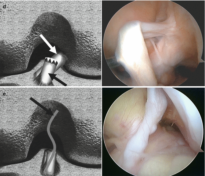
Fig. 28.1
Classification of the ACL remnant [21]. (a) Type 1: ACL remnant (black arrow) bridging the PCL (white arrow) and tibia. Normal attachment of the ACL to the femur was entirely lost. (b) Type 2: ACL remnant (arrow) bridging between the intercondylar notch and tibia. Normal attachment of the ACL to the femur was entirely lost. (c) Type 3: partial rupture of the posterolateral bundle (black arrow). The anteromedial bundle (white arrow) of the ACL has an attachment of femoral origin and is well preserved. (d) Type 4: partial rupture of the anteromedial bundle (black arrow). The posterolateral bundle (white arrow) of the ACL has an attachment of femoral origin and is well preserved. (e) Type 5: no substantial ACL remnants bridging the tibia and either the femur or PCL. The diameter of the proximal ACL remnant is attenuated to less than one-third of its original size, and the tension of the remnant is accordingly loose (arrow)
Colombet and Dejour et al. performed arthroscopic evaluation of the ACL rupture (a continuous series of 418 patients) [6]. They also included partial ACL tear in their classification. The ACL remnants were divided into four categories: totally disappeared ACL (50 %), posterolateral bundle conservation (16 %), healing on PCL (23 %), and anteromedial bundle conservation (11 %). In addition to the above classification, Dejour et al. subdivided the groups of partial ACL injury [5]. In the case of a partial ACL injury, further dynamic evaluation of the mechanical integrity of the remaining bundle was performed by palpation with a probe. The remaining bundle was classified as functional or nonfunctional, depending on the presence of mechanically solid fibers or the ability of the examiner to further stretch them, respectively. If the remaining bundle resisted further stretching, they were classified as functional. If the remaining bundle was lax and the examiner could stretch them significantly further, they were classified as nonfunctional. The authors found a significant difference between the occurrence of a functional remnant in the PL-intact group (functional, 67 %; nonfunctional, 33 %) and the occurrence of functional remaining fibers in the AM-intact and PCL-healing groups (functional, 17 %; nonfunctional, 83 %).
In order to determine the treatment strategy, Kazusa and Ochi et al. [34] classified the ACL remnant pattern as follows. Group 1 is partial rupture of the ACL (Group 1a, partial rupture of the PL bundle; Group 1b, partial rupture of the AM bundle; and Group 1c, partial rupture of the ACL but the remaining bundle could not be ascribed to either the AM or PL bundles). Group 2 is complete rupture of the ACL (Group 2a, ACL remnant bridging the PCL and tibia; Group 2b, ACL remnant bridging the roof of the intercondylar notch and tibia; Group 2c, ACL remnant bridging the lateral wall of the intercondylar notch and tibia; and Group 2d, no substantial ACL remnants bridging the tibia and either the femur or the PCL). Group 1a and Group 1b are indications for the single-bundle ACL augmentation. Group 1c, 2a, 2b, and 2c are indications for the single- or double-bundle ACL augmentation. Currently, we do not perform ACL augmentation for Group 2a.
In summary, although the diagnosis of a partial versus a complete ACL tear can be made with greater accuracy during arthroscopy, the decision as to whether the ACL remnant is preserved and ACL augmentation performed should be made after thorough consideration of clinical tests, laxity measurements, MRI, and arthroscopic findings.
References
1.
2.
3.
4.
5.
6.
7.
8.
Ochi M, Adachi N, Deie M, Kanaya A (2006) Anterior cruciate ligament augmentation procedure with a 1-incision technique: anteromedial bundle or posterolateral bundle reconstruction. Arthroscopy 22:463.e1–e5Crossref
9.
Ochi M, Adachi N, Uchio Y, Deie M, Kumahashi N, Ishikawa M, Sera S (2009) A minimum 2-year follow-up after selective anteromedial or posterolateral bundle anterior cruciate ligament reconstruction. Arthroscopy 25:117–122CrossrefPubMed
Stay updated, free articles. Join our Telegram channel

Full access? Get Clinical Tree


