Deltoid Injuries


Deltoid Injuries


Jerry S. Sher

Joseph P. Iannotti


J. S. Sher: Clinical Assistant Professor, Department of Orthopaedics and Rehabilitation, University of Miami School of Medicine, Miami Beach, Florida.

J. P. Iannotti: Professor and Chairman, Department of Orthopaedic Surgery, The Cleveland Clinic Foundation, Cleveland Clinic Lerner Foundation, Cleveland Clinic Lerner College of Medicine, Cleveland, Ohio.


INTRODUCTION

Injuries to the deltoid muscle occur relatively infrequently compared with other, more common shoulder disorders such as those involving the rotator cuff. Regardless of the cause, these injuries can often result in significant shoulder dysfunction. An understanding of the functional anatomy of the deltoid and the mechanisms of injury can help to minimize iatrogenic causes and enable the formulation of optimal treatment strategies.


ANATOMY AND FUNCTION

The importance of the deltoid in providing active elevation of the arm is readily appreciated when considering the compromised function associated with pathologic conditions affecting this muscle. Approximately 60% of the strength in abduction and elevation in both the coronal and scapular planes, respectively, is attributed to the deltoid, as determined after anesthetic block of the axillary nerve. Progressive loss of muscle force with increased abduction and early fatigue further compromises shoulder function in such individuals.16

Some reports cite sporadic cases of functional elevation of the arm in patients with isolated and complete paralysis of the deltoid.3,19,36,58 Preservation of active elevation was attributed to the compensatory action of the rotator cuff, pectoralis major, trapezius, and serratus anterior muscles.3 Despite the ability to raise the unweighted arm, considerable deficits in abduction strength occurred in addition to abnormal scapulohumeral mechanics, impairment of humeral extension, and early fatigability.3,19,58

Anatomically, the deltoid comprises three distinct heads: the anterior, the middle, and the posterior portions, which vary in both structure and function. Its extensive origin forms a horseshoe-type configuration arising from the distal one-third of the clavicle, the acromion, and the lateral one-third of the spine of the scapula. Distally, the muscle converges to insert on the deltoid tuberosity of the middiaphysis of the humerus. Its broad origin, which is derived from the mobile scapula and clavicle, affords the deltoid a mechanical advantage by allowing the muscle to maintain its resting length at various arm positions. The bipennate structure of the large middle head contributes to abduction strength through contraction of its fibers at an angle to the line of pull, which also serves to maintain muscle fiber resting length and improve efficiency. In contrast, muscles with a parallel fiber arrangement such as the anterior and posterior deltoid, by virtue of their structural configuration, result in considerably decreased strength during contraction.21

Differences in activity of the deltoid’s three portions, relative to arm position, have also been observed through electromyographic analysis.57 The anterior and middle heads remain active at all angles of abduction and in multiple planes (coronal, scapular, and parasagittal), whereas the posterior deltoid, also an important shoulder extensor, contributes to elevation when the arm is above 110 degrees.57 Abduction in the coronal rather than scapular plane effects a relative increase in posterior deltoid function and a decrease in anterior deltoid activity.31 When the arm is in abduction, the posterior deltoid functions as a secondary external rotator, and its clinical importance is increased in patients with massive rotator cuff tears involving the infraspinatus tendon. Its function as a secondary external rotator is greatest with the arm posterior to the plane of the scapula and at 90 degrees of abduction.


Neural innervation is afforded by the axillary nerve, which takes a circuitous path before entering the deltoid muscle. It arises from the posterior cord of the brachial plexus and courses across the inferolateral border of the subscapularis approximately 3 to 5 mm medial to the musculotendinous junction.37 It passes inferior to the glenohumeral axillary recess and exits the quadrangular space where it divides into two trunks. The posterior trunk splits and innervates the teres minor and posterior deltoid before terminating as the superior lateral cutaneous nerve. The anterior trunk winds around the humerus and innervates the remaining deltoid muscle. It becomes subfascial and intramuscular at a point between the anterior and middle heads.13 It is generally located one-third the distance from the lateral acromion to the deltoid tuberosity, which in most patients is 4.4 cm from the lateral acromion.


TABLE 34-1 CAUSES OF DELTOID INJURIES


Cause


Nerve


Muscle


Iatrogenic


Iatrogenic


Incomplete injury


Detachment of origin


Complete injury (transection, traction, compression)


Soft tissue


Lateral acromionectomy


Trauma


Complete acromionectomy


Glenohumeral dislocation


Infection


Fracture


Necrosis/fibrosis


Blunt trauma


Posttraumatic injury


Penetrating trauma


Fracture/dislocation


Entrapment


Blunt


Quadrilateral space syndrome


Penetrating


Cervical radiculopathy


Contracture


Brachial plexopathy


Inflammatory


Infection


Polymyositis


Upper motor neuron disease


Dermatomyositis


Lower motor neuron disease


Infection


Myoneural junction (myasthenia gravis)


Candida


Mycoplasma


Trichinosis


Toxoplasmosis


Viral


Other


Metabolic/endocrine


Hypothyroidism


Acromegaly


Other


Steroid myopathy


Alcoholic myopathy


Electrolyte imbalance (hypokalemia)


Muscular dystrophy


Amyloidosis


Spontaneous detachment of the deltoid origin (chronic massive rotator cuff tear)


Cause

Although a host of disorders can affect the function of the deltoid, this chapter focuses on lesions isolated to the axillary nerve and those affecting the musculotendinous integrity of the deltoid. For the purpose of completeness, Table 34-1 has been included and represents a comprehensive list of causative factors that can contribute to deltoid muscle dysfunction.


DETACHMENT OF THE DELTOID ORIGIN


Cause

Surgical techniques that use open methods of rotator cuff repair and acromioplasty often require detachment of a portion of the deltoid origin to facilitate exposure.
Although many patients demonstrate an uneventful postoperative course, a few develop complications related to dehiscence of the deltoid repair, which can manifest as a residual defect at the muscle’s origin (Fig. 34-1). Similar but less frequent occurrences have been reported after deltopectoral, deltoid splitting, and axillary surgical approaches for instability, degenerative, and traumatic conditions of the shoulder.25 Factors thought to contribute to this postoperative complication include inadequate surgical repair of the deltoid origin, osteoporotic bone unable to withstand the tension afforded by transosseous sutures, poor tissue quality, overzealous retraction, and patient noncompliance. This outcome is avoidable in most cases, but when it does occur, it is typically associated with considerable morbidity and disability.6,20,25,32,45,46,55,56,64


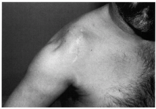
Figure 34-1 Shoulder with middle deltoid detachment 2 years after anterior acromioplasty and rotator cuff repair.

Recent reports have implicated arthroscopic acromioplasty as a potential cause of detachment of the deltoid origin (Fig. 34-2).10,34,59 In an anatomic study, Torpey and colleagues59 noted that the anterior and anterolateral acromion have a direct tendinous attachment of the deltoid muscle, whereas the muscle attaches to the dorsal side of the acromion through a periosteal attachment. Mathematical models indicate that an anterior inferior acromioplasty of 4 and 6 mm would release 41% and 69% of the tendinous origin, respectively.16,59 Another report10 confirms detachment of the deltoid origin during an arthroscopic subacromial decompression and highlights this complication, which is likely underreported. Although most patients do satisfactorily after arthroscopic acromioplasty, caution is warranted in performing aggressive bone resection. Moreover, although the anterior deltoid origin may be weakened after this procedure, superior fibers remain in continuity preserving deltoid function.


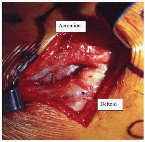
Figure 34-2 Chronic defect in the anterior deltoid origin, seen at the time of revision open repair, secondary to an excessive detachment of the deltoid origin during an arthroscopic acromioplasty.


Atraumatic or Spontaneous Deltoid Origin Detachment

Sporadic cases of spontaneous rupture of the deltoid origin have been described in the literature.7,43,48 The senior author has described a deltoid origin rupture in the absence of any iatrogenic event in four shoulders of three patients.7 All cases were associated with chronic massive tears of the rotator cuff and cephalad migration of the humeral head (Fig. 34-3). Poor shoulder function was evident throughout, and a maximum of 30 degrees of arm elevation was evident in one patient. Notably, weakness rather than pain was the predominant symptom. Another report cites two cases of spontaneous deltoid rupture involving the middle head of the muscle.43 Both cases also had concomitant massive rotator cuff tears.

Some common findings included advanced age more than 60 years in all cases and involvement of the middle head of the deltoid in most cases. Repeated cortisone injections are also believed to play a causative role in some instances. One proposed explanation for spontaneous rupture includes increased friction or mechanical impingement of the greater tuberosity against the proximal deltoid. Absence of a functional rotator cuff can cause cephalad migration of the humeral head with resultant abnormal or excessive contact between the deltoid and greater tuberosity (Fig. 34-4). Chronic mechanical deltoid impingement, decreased muscle mass, and repeated corticosteroid injections may all contribute to this uncommon occurrence. These cases highlight that a new onset of shoulder weakness in the presence of an existing massive rotator cuff tear may be secondary to deltoid rupture rather than extension of the rotator cuff tear.


Diagnosis

Assessment of an affected patient should include consideration of the individual’s overall complaints, precipitating factors, functional status, and associated pathologic conditions. Evaluation may reveal symptomatic arthritis of the glenohumeral or acromioclavicular joints, failure of rotator cuff repair, inadequate acromioplasty, excessive acromion
resection (acromionectomy), or neurologic impairment in association with deltoid detachment. Pain at the anterolateral aspect of the shoulder, for example, could be related to a refractory rotator cuff disorder, disruption of the deltoid origin, or both. This may also hold true for a patient’s inability to elevate the arm. Evaluation may reveal functional deficits secondary to rotator cuff dysfunction, deltoid dysfunction, or both.


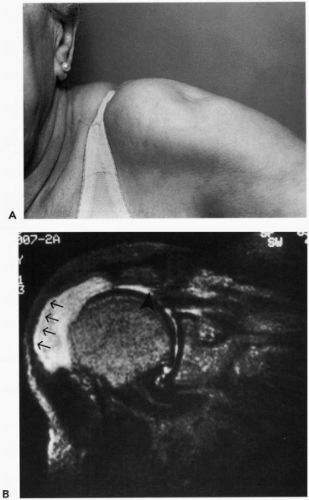
Figure 34-3 (A) Spontaneous middle deltoid origin detachment in a patient with a massive chronic rotator cuff tear. (B) Magnetic resonance scan demonstrates the deltoid origin disruption (arrows) and rotator cuff tear (arrowhead).


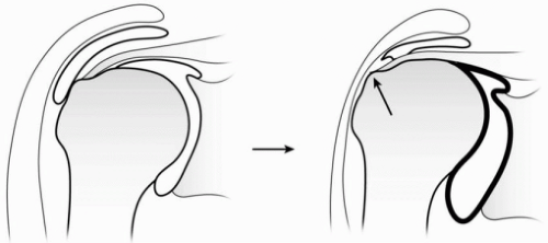
Figure 34-4 Mechanism of wear on the undersurface of the deltoid muscle in a shoulder without (left) and with (right) a rotator cuff tear. A deficient rotator cuff allows for cephalad migration of the humeral head with chronic abrasion of the greater tuberosity against the deltoid muscle. (Reproduced with permission from Morisawa K, Yamashita K, Asami A, et al. Spontaneous rupture of the deltoid associated with massive tearing of the rotator cuff. J Shoulder Elbow Surg 1997;6:556.)


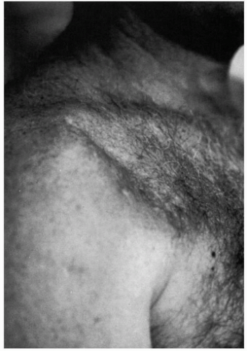
Figure 34-5 Postoperative anterior deltoid detachment.

Patients with disruption and retraction of the deltoid origin frequently display a defect and a loss of normal muscle contour at the sight of detachment with enlargement (bulge) of the deltoid distal to the site (Fig. 34-5). This deformity is best seen with the arm in abduction against gravity or resistance. The deformity may be difficult to see in obese patients. Smaller deficiencies may not be readily apparent with
the arm at the side, but they can often be appreciated when the humerus is actively abducted. Palpation along the acromion border can facilitate assessment of the size of the defect and the degree of retraction. Frequently, focal tenderness can be elicited at the detachment site. Atrophy of the deltoid or rotator cuff may also be evident and can represent disuse, muscle or tendon injury, or neurologic impairment.

Although a thorough history and examination can reliably generate a diagnosis, additional studies are often helpful in excluding alternate sources of shoulder pain. Plain radiographs can facilitate the assessment of acromion morphology, the adequacy of decompression, and the presence of a prior acromionectomy (Fig. 34-6). Diagnoses of glenohumeral and acromioclavicular joint arthritis or cervical spondylosis may be realized. Cephalad migration of the humeral head can confirm any chronic tearing or dysfunction of the rotator cuff. Electrodiagnostic studies can provide objective confirmation of neurologic dysfunction and are generally indicated to rule out deltoid denervation. Arthrograms and magnetic resonance images can aid in the evaluation of rotator cuff integrity. Magnetic resonance imaging can also demonstrate deltoid detachment. It helps define the degree of muscle atrophy and retraction that can correlate with the ability to achieve a successful primary repair (Fig. 34-7).


Functional Outcome

Many investigators have confirmed the overall poor results of rotator cuff repair when the abnormality was associated with detachment of the deltoid origin.6,20,25,45,46,55,56,64 In some cases, function is satisfactory although not normal (Fig. 34-8). In these patients, the deltoid detachment is often small, and there is evidence of intact rotator cuff with minimal pain. Bigliani and colleagues6 reported a series of 31 patients who underwent reoperation for failed rotator cuff repairs. At follow-up, 15 individuals had unsatisfactory results, and 9 of these demonstrated associated deformities of the deltoid origin. Overall, 13 of the 31 individuals had deltoid detachments.6 Neviaser and Neviaser46 published the results of 50 patients who underwent repeated surgery for failed repairs of the rotator cuff. Six of 17 patients who had less than 90 degrees of active elevation before reoperation still had poor active motion (<90 degrees) postoperatively. All 6 of the patients had deltoid deficiencies. Closer analysis of this group revealed that 4 had irreparable detachments of the deltoid origin, 1 had deltoid fibrosis as a result of a previous postoperative infection, and 1 had a permanent axillary nerve palsy.46


Figure 34-6 Lateral acromionectomy in a patient with a middle deltoid detachment.


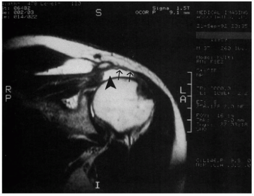
Figure 34-7 Postoperative magnetic resonance image demonstrates a detachment of the middle deltoid (arrows) and full-thickness tear of the supraspinatus tendon (arrowhead).

Only gold members can continue reading. Log In or Register to continue

Stay updated, free articles. Join our Telegram channel

Jul 15, 2016 | Posted by in ORTHOPEDIC | Comments Off on Deltoid Injuries

Full access? Get Clinical Tree

Get Clinical Tree app for offline access