Fig. 12.1
3D reconstruction CT is important in order to visualise osteophyte distribution and assess complex deformity patterns when planning surgical debridement
The accuracy of the method of diagnostic imaging of the soft tissue structures around the elbow continues to raise considerable debate. MRI can be useful to diagnose collateral ligament injuries but also in the evaluation of loose bodies, osteochondral lesions, olecranon osteophytes and neurologic complaints [11].
12.3 Management in Degenerative Elbow
The mainstay of early treatment of the young patient with posttraumatic arthritis consists of maintaining joint mobility and reducing activities that place stress across the elbow such as weight-bearing or repetitive motions. Nonsteroidal anti-inflammatory drugs and selective intraarticular corticosteroid injections can control pain and facilitate daily use of the degenerative elbow.
After considering 6-month failure of conservative treatment (mobilisation, splinting and physical therapy), intact articular space, absence or mild anatomical incongruency, ROM reduction and sport- and occupation-related disability, a patient can be a candidate for an arthroscopic arthrolysis [21, 28]. On the other hand, arthroscopic technique can be useful in association with open surgery in order to avoid large surgical approaches. Sometimes removal of a columnar plate or screws can be associated with an arthroscopic arthrolysis.
12.4 Osteochondritis Dissecans Lesions
Osteochondritis dissecans occurs most commonly in overhead-throwing athletes and in gymnasts between the ages of 13 and 16 years [8, 12]. It typically affects the young adolescent athlete involved in high-demand, repetitive overhead or weight-bearing activities. The most commonly associated sports are baseball, gymnastics, racquet sports, football and weightlifting [3, 8].
OCD can be a cause of painful elbow with limited ROM. These young patients, usually athletes complaining pain and dysfunction, limit their activity becoming unable to participate in sport. Although lesions have been reported in the trochlea, radial head and olecranon, the most common site of OCD of the elbow is in the capitellum [3, 8, 12].
Radiographs reveal rarefaction, radiolucency or fragmentation of the anterolateral capitellum. MRI has become the standard imaging modality to identify OCD, and it can provide an accurate assessment of the size, extent and stability of the lesion.
Determination of lesion stability and the integrity of the articular cartilage cap are really important regarding the decision to prescribe nonoperative treatment or proceed with the surgery [8].
Panner’s disease, most common between 4 and 8 years of age, should not be confused with true OCD because it involves the entire ossification centre, while only the anterolateral capitellum is involved in osteochondritis dissecans of the capitellum [3, 8].
Treatment for stable, early stage OCD lesions consists in avoiding repetitive stress of the elbow and observation. If the lesion has not resolved in 3–6 months, then consideration of surgical management is made.
Surgical management is the treatment of choice for unstable lesions, lesions that have failed nonoperative management and loose bodies. Lesions that are unstable have a tendency to remain symptomatic even if no loose body is present, therefore leading to surgery [8].
Multiple operative procedures have been described for treating OCD. Surgical treatments include drilling of the lesion, fragment removal with or without curettage of the residual defect, fragment fixation by a variety of methods (pull-out wiring, Herbert’s screw, bone peg grafting, etc.), reconstruction with osteochondral autograft and autologous chondrocyte implantation [3].
In the literature, several studies report different results with open procedure, but more recently arthroscopy has been employed with encouraging scores in the treatment of capitellar OCD [3, 8, 22].
Baumgarten and colleagues report excellent results in a group of 17 patients whose elbows were treated with arthroscopic debridement with a complete return to sport activities at the pre-injury level in 82 % of cases [3, 5, 8].
Reports of arthroscopic treatment of OCD of the capitellum with removal of loose bodies, debridement and abrasion chondroplasty describe overall improvements in pain and range of motions with variable return to pre-injury level of sporting activity [3, 22].
A grading system based on absent, partial or total detachment of the bone plug has been developed by Baumgarten et al. to aid in decision-making during elbow arthroscopy. The recommendation presented for Grade 1 lesions is either observation or arthroscopic drilling of the lesion. Grade 2 lesions were treated with debridement of the cartilage to healthy tissue. Grade 3 lesions were treated with loosening of the fragment to create a Grade 4 lesion, which was then resected. Grade 5 lesions were treated with a diligent search for the loose bodies [5, 8].
In our patients we prefer arthroscopic evaluation and treatment for lesions requiring operative management. Removal of the bone plug and microfracture is mandatory in order to eliminate catching and popping, while it is still controversial the possibility to bone graft the lesion [22].
In some cases we have been performed an arthroscopic mosaicplasty taking the graft from the homolateral knee putting the patient in lateral decubitus and extrarotating the hip performing knee arthroscopy. The 6.5 mm cylinder graft token from the lateral knee trochlea was inserted in the elbow lesioned area carefully checking the angle of the drilling and of the insertion of the bony cartilaginous cylinder. Arthroscopically the perpendicular insertion of the cylinder allows a complete coverage of the OCD area. Four months later MRI shows a nice bone incorporation of the graft. Post-operatively the CPM started in day 2 and passive exercises in day 4 post-op. Patients were back to normal activity in 4 months [22] (Figs. 12.2, 12.3 and 12.4).
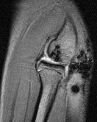
Fig. 12.2
The mosaicplasty from the knee to the elbow is performed on lateral decubitus positioning and the hip in extrarotation to allow knee arthroscopy for taking the graft from the lateral trochlea
Fig. 12.3
The graft is positioned on the lateral humeral condyle to fill the OCD gap
Fig. 12.4
Four-month control MRI shows a good bone incorporation of the osteochondral cylinder
12.5 Arthroscopic Technique in Degenerative Elbow
Arthroscopy is increasingly used to diagnose and treat elbow pathologies although the elbow has always been considered a difficult joint to be endoscopically explored. Arthroscopy knowledge increase and technology breakthrough in the last few years have allowed a standardisation of techniques and a better definition of indications. In the 1980s, Andrews J. R. and Carson W, G, [2], Hempfling H. [10] and Lindenfeld T. N. [15] published the first indications, techniques and notions on elbow arthroscopy. In 1981, on the basis of their observations, Morrey et al. determined that the elbow functional motion ranged from 30 to 130° of flexion [17]; however, a lot of daily activities performed at work or while doing physical exercise require an extension above 30°. As a matter of fact, for sportsmen and manual workers even a small decrease in ROM, together with slight symptoms of pain and inability to perform specific tasks, can be unacceptable and, hence, interfere with their daily work or sport activities. For this reason, there has been an extension of indications concerning stiff elbows treatment [16].
In 1992, O’Driscoll and Morrey presented 72 cases of elbow arthroscopy [19], and in 2001 they published a review of 473 cases in which they analysed the complications related to this procedure [13]. In the previous year, Reddy A. S. et al. [24] had published a review of 172 cases in which patients had undergone arthroscopic elbow surgery with a 7-year follow-up. The list of indications for elbow arthroscopy has grown over the past years, and today it includes osteochondritis dissecans (OCD), plica syndrome, synovectomy in R.A. and other synovitis, lateral epicondylitis, loose bodies removal [23, 29] and stiff elbows related to degenerative or posttraumatic causes [6, 16]. Recently, Conso C. [9] as well as Shubert T. [27] and Salini V. [26] have published the results obtained by comparing, respectively, 32, 24 and 15 arthroscopic cases presenting a moderate stiffness of the elbow and other pathologies, with those obtained with open techniques. There are several studies regarding this subject in the literature, but all of them are based on a small number of patients with a variety of pathologies treated with different surgical techniques [1, 4, 18, 20, 25, 30].
Anaesthetist identifies nerve trunks by applying electrostimulation and places a catheter without injecting the anaesthetic. Patients then undergo general anaesthesia. When they wake up, only after a neurological evaluation, peripheral block is performed. After the induction of anaesthesia, ROM is carefully assessed and a complete ligamentous balancing is carried out. The tourniquet is inflated to 250 mmHg. The patient is then placed prone, with the shoulder abducted 90°, the elbow flexed to 90° and the arm held up by an arm holder secured to the operating table. Sterile field is set up and elbow joint landmarks are drawn by a dermographic pen (medial and lateral epicondyle, ulnar nerve, radial head, posterior soft spot). Soft spot posterior portals, supero-antero medial portals and supero-antero lateral portals are marked. Ulnar nerve neurolysis has always been performed by making a 2 cm skin incision, except in full ROM cases (full ROM painful elbows, occasionally decreased ROM). An 18-gauge needle is inserted in the elbow through the “soft spot” in the middle of the triangular area delimited by the epicondyle, the radial head and the olecranon, while the joint is distended by injecting 20 ml of normal saline solution to introduce the trocar while shifting neurovascular anterior structures away. Five portals, three posterior and two anterior, are always used. After the incision is made, soft tissues are retracted by using a fine haemostat. Posterior compartment arthroscopy is firstly performed by introducing a 4,5 mm 30° arthroscope through the posterolateral portal (soft spot). Then a second portal is established, 1,5 cm proximal to the latter. These two portals allow to use the scope and the shaver at the same level of the posterior portion of the radial head. Joint distension is achieved by a pump set at 35–50 mmHg. Once we get a good and complete view of the proximal radio-ulnar joint (posteriorly), a third posterior portal is placed in the olecranon fossa, close to the triceps medial border and oriented 2–3 cm proximal to the olecranon tip. A complete olecranon fossa and its lateral wall debridement can be performed as well as, if present, a lateral olecranon and humerus loose bodies removal to allow a better sliding of the articular surfaces. We use a different approach related to osteophyte dimension and ulnar nerve presence on the medial side. After inserting the arthroscope through the most proximal portal, we evaluate osteophyte dimensions; if they are small, we protect the ulnar nerve by positioning a retractor in an accessory portal slightly posterior to the ulnar nerve, and we resect the osteophytes arthroscopically. If they are large, we prefer to remove the osteophytes by performing a small arthrotomy at the end of the procedure, thus avoiding fluid extravasation during arthroscopy. The medial approach is always used after ulnar nerve neurolysis, which is the first surgical step of the procedure. This is necessary to prevent the overstretching of the nerve testing flexion and extension during elbow arthroscopy. The scope is then introduced in the anterior compartment through the supero-antero medial portal, 2 cm proximal and 1 cm anterior to the epitrochlea. In the stiff elbow the medial approach is preferable because it allows to locate the ulnar nerve by palpation, which is not possible on the lateral side. The anterolateral portal is created using an inside-out technique and placing a Wissinger rod 2 cm proximal and 1 cm anterior to the lateral epicondyle. A plastic cannula is introduced on the rod, and, subsequently after having the rod removed, a shaver can be positioned and the anterior debridement carried out (removal of loose bodies, anterior osteophytes and synovectomy). In several cases, in the presence of a thick capsule (posttraumatic causes), an anterior capsulectomy may be required. We start trimming the proximal humeral capsule by a shaver, but the real anterior capsulectomy is performed by a basket forceps, at about 1 cm proximally to the apex of the coronoid, firstly in a lateral-medial and then in a medial-lateral direction. After arthroscopy, ROM is assessed. One or two suction drainages are positioned into the joint, arthroscopic accesses are sutured and a splint holding the joint in full extension is applied to correct the articular loss of extension. On post-op day 1, patients start a 20 min continuous passive motion (CPM) four times a day, together with an assisted physiokinesis therapy, at least 60 min per day. On day 2 they start a self-assisted active and passive mobilisation in flexion-extension. On day 3 drains are removed and we continue with the rehabilitative programme. Indomethacin 50 mg three times per day is somministrated for 15 days. At the time of discharge from the hospital, patients are taught of the exercises they need to practise at home. They continue the same programme with a therapist for 3 months.
Related posts:


Stay updated, free articles. Join our Telegram channel

Full access? Get Clinical Tree


