, Paul D. Siney1 and Patricia A. Fleming1
(1)
The John Charnley Research Institute Wrightington Hospital, Wigan, Lancashire, UK
Deep Infection [1]
We define deep infection as – infection around the implant.
The most likely source of infection that was not under control by standard aseptic precautions appeared to be the air of the operating room. It was therefore decided to build a clean air operating enclosure.
“The general opinion would seem to be that the air of an average operating theatre is relatively innocuous. In the ward, on the other hand, the aerial route for post–operative cross–infection is regarded as potent…” “This was the opinion I myself held until some two years ago when a very high rate of wound infection in a new type of arthroplasty prompted this study” (1964).
Post–operative sepsis, resulting in the infection around the implant, during the years 1958–1967, was so high (8.9 %) that it would have been necessary to abandon this type of surgery had not special precautions in the operating theatre shown a marked improvement.
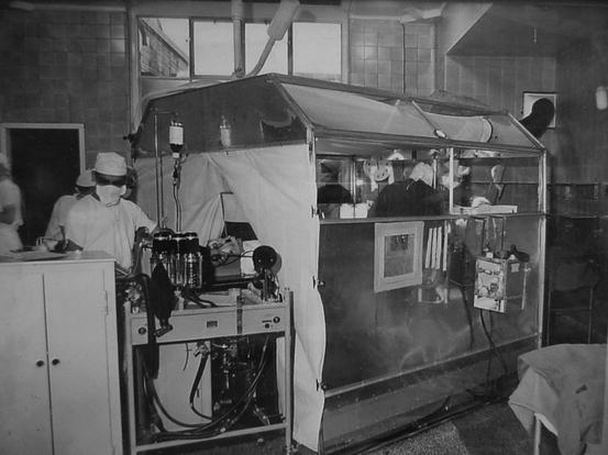
A prototype filtered air enclosure was constructed to contain the lower half of the patient’s body and the three surgeons of the operating team. Filtered air is forced in at the top of the enclosure and the surgeons wear respirators through which their exhaled air is extracted so as not to mix with the filtered air of the enclosure. (1964) (Fig 13.1).
Fig. 13.1
The original clean air enclosure “the Greenhouse” at Wrightington Hospital
The First Survey
The study related to 2170 consecutive arthroplasties of the hip joint performed between January 1959 and September 1967 (Table 13.1, Fig. 13.2) “… to permit at least 12 months to elapse between the last operation and completing the report.”
Table 13.1
Study of infection after low-frictional torque arthoplasties – Jan 1959 to Sept 1967 and subsequently (Fig. 13.2)
Phase | Period | Operating room | Colonies per plate per hour | No. of hip replacements | Infection rate % |
|---|---|---|---|---|---|
1 | Jan 1959 to Nov 1961 | Primitive | 80–90 | 190 | 8.9 |
2 | Nov 1961 to June 1962 | Prototype filtered air enclosure 10 air changes per hour | 2.5 | 108 | 3.7 |
3 | June 1962 to March 1966 | Clean air enclosure 130 air changes per hour | 1.9 | 1164 | 2.2 |
4 | June 1966 to Sept. 1967 | Clean air enclosure 300 air changes per hour | Air not sterile | 708 | 1.3 |
Lowest that can be recorded | |||||
5 | 1969 – | As above | Lowest recorded | 1000 | 1.0 |
Total body exhaust suits |
Fig. 13.2
Graph of infection rates. The study related to 2170 consecutive arthroplasties of the hip joint performed between January 1959 and September 1967. Further information was added to 1974 (Reproduced from Wroblewski [10])
“It was not possible to exclude a number of additional variables which could contribute to a reduction in wound infection … improved form of wound closure … the bodies of the surgeons or the others in the operating team, through permeability of the textile gowns.” “Though the filtered air enclosure has improved the air cleanliness 25 times … it is still not completely sterile. It would be unlikely that a total abolition of infection could be expected.” “Despite extreme precautions our rate is still about 1.8 %” (1968).
It was in the early stages of the development of the clean air operating enclosure (1964) “… another line … A form of ‘air curtain’ in which the enclosure acts as a hood or shield to reduce the tendency of the cold air flow to entrain infected particles.” Charnley was clearly considering the next stage in the development of clean air enclosure – without side panels, pointing out that this “… will effect an economy in the volume of air required and necessitate in the method of illumination.”
The complexity of the problem becomes obvious when one considers numbers of operations, diagnostic criteria, follow-up, and the time interval from the primary operation to establishing the diagnosis of deep infection.
Establishing the Diagnosis
Clinical experience suggests that establishing the diagnosis of deep infection is easier in cases referred for a second opinion than after our own operations.
Clinical Diagnosis
Pain relief after THA is such a consistent feature that failure to relieve pain must put the surgeon on guard. Barring any problems at surgery there are only three possibilities: inappropriate indication, inappropriate patient selection, infection.
Inappropriate Indication
Clinical assessment fails to identify the source of pain. The radiograph is viewed before, or at the same time, as taking the history and examining the patient. Clinical assessment must come before viewing radiographs; radiographs merely confirm and offer a record of what is usually obvious from the history and examination.
Inappropriate Patient Selection
It must not be assumed that every patient with an arthritic hip would benefit from THA. Rare though it may, beware of a patient with a very high expectation and flattering the surgeon’s apparent skill and reputation. Does the surgeon’s ability match patient’s expectations? It must not be assumed that to do something is to do good, while to advise delaying surgery implies unwillingness to help, or negligence.
Complications
Dislocation is obvious, loosening of components comes later and is largely asymptomatic, fracture of the stem is now a late rarity – that leaves infection.
History of haematoma, delayed wound healing, courses of antibiotics – increase the index of suspicion. Regular follow-up with good quality radiographs and continuity of observer method is essential.
Bacteriology in the study of deep infection after THA is outside the scope of this work. It is best left in the hands of the experts on the subject.
Classification of Deep Infection
The practical usefulness of any classification is inversely proportional to its complexity. Infection will continue to be studied retrospectively if meaningful information is to be gathered.
Early Infection
Signs of inflammation, delayed would healing, infected haematoma, purulent discharge and early sinus formation. Wound dehiscence of deep fascia is extremely rare. Early exploration under conditions as in the primary operation, evacuation, examination of the deep fascia for deep extension, but without probing or opening unless communication is obvious.
Charnley arranged for photographs of the wounds to be taken before discharge from the hospital: 5400 photographs are available for further studies to anyone interested in the subject.
Late diagnosis of early infection is probably the most common scenario. Delays or lack of follow-up, inadequate radiographs, and lack of awareness of the possibility of infection.
Late presentation following early contamination. This is a complex subject that continues to be debated. If this is a true entity in clinical practice then fortunately it has not presented itself, as would have been expected, with increasing follow-up.
Late haematogenous infection – fortunately very rare. The criteria for the diagnosis must be strict: Primary surgery in a patient not at risk, no post-operative complications suggesting a possibility of infection. Clinical success with normal radiographs – well documented over a period of probably not <3 years, a source of possible bacteraemia, for example lower limb skin infection.
Related posts:


Stay updated, free articles. Join our Telegram channel

Full access? Get Clinical Tree


