6 Cortical Screw Systems
6.1 Introduction
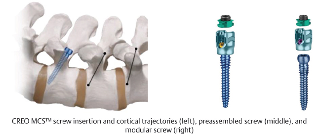
The posterior approach for cortical screw fixation is described in Chapter 3 Posterior Retractor Systems. Posterior screw fixation with a cortical trajectory was initially described by Santoni et al in 2009.1 Cortical screws were developed with the intention of maximizing cortical bone purchase, which has proven advantageous in conditions such as osteoporosis where trabecular bone may not have adequate density to achieve stable fixation.2 Regarding placement, the cortical screw start point is located at the junction of the superior articular process and pars interarticularis.3 Utilization of this start point allows for intraoperative preservation of muscle attachments, and possible reduced postoperative pain.3,4,5 The trajectory of cortical screws is lateral and cephalad from the start point.6 This trajectory allows for placement of the screw within the cortical bone of the pedicle, rather than entering the trabecular bone of the vertebral body as is the case with pedicle screws. Minimally invasive cortical screw placement can be utilized during midline lumbar fusion (MIDLF), which involves a posterior midline approach and microsurgical laminectomy, followed by cortical screw fixation.7,8 This technique reduces the risk of neural injury due to the medial to lateral and caudal to cephalad trajectory of the cortical screw (▶ Fig. 6.1).7 Additionally, MIDLF provides for decompression and fusion through the same surgical corridor, further reducing tissue dissection and retraction.7
6.1.1 Components
The dimensions of cortical screws necessarily differ from those of pedicle screws. Cortical screws have a denser thread, larger minor diameter, and a smaller bite than traditional cancellous pedicle screws (▶ Fig. 6.2).9 The denser thread and amount of depth per rotation aid insertion into harder cortical bone. Cortical screws are also shorter than pedicle screws, so as not to violate the pedicle borders or enter the trabecular bone of the vertebral body. Similar to pedicle screws, the heads of cortical screws contain articulating tulips that allow for percutaneous placement of rods to promote stable fixation of two vertebral levels.
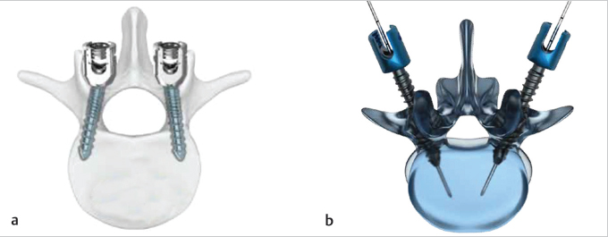
Fig. 6.1 Screw trajectories for (a) cortical screws (Arsenal CBx) and (b) pedicle screws (Illico MIS posterior fixation).
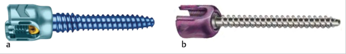
Fig. 6.2 Dimension differences between (a) cortical (CREO MCS, Globus Medical) and (b) pedicle screws (SpheRx DBR III, NuVasive, Inc.).
6.1.2 Efficacy and Complications
The effectiveness of cortical screw fixation has been extensively studied from a biomechanical perspective. Many biomechanical studies utilizing cadaveric spine constructs have demonstrated that cortical screws are at least equivalent to pedicle screws in regard to bone purchase.2,9,10,11,12,13 Regarding strength, multiple studies have indicated that cortical screws have a superior strength profile compared to pedicle screws. Cortical screws have demonstrated increased resistance to craniocaudal toggling forces, greater insertional torque, and a greater pullout load required before screw failure occurs. The increased strength of cortical screws is attributed to the cortical, rather than trabecular, trajectory of placement within the pedicle.
The study of peri- and postoperative outcomes after cortical screw fixation is an emerging topic in the literature, with recent data providing promising results. Studies have demonstrated reduced surgical morbidity with the use of cortical screws compared to pedicle screws.3,4,5,8 Cortical screw use has been associated with shorter incision lengths, shorter operative times, reduced intraoperative blood loss, and reduced inpatient postoperative pain. These improvements have been attributed to minimization of soft-tissue distraction and retention afforded by the more medial screw start point. Longer-term postoperative outcomes, however, have been mixed.3,5 Some studies have demonstrated similar pain, functional status, and fusion rates at 12 months postoperatively in cohorts receiving either cortical or pedicle screws.5 Other studies have noted an increased pain profile in cortical screw cohorts at 6 to 8 months postoperatively.3 Further prospective, controlled studies are required to better elucidate the long-term clinical and radiographic efficacy of cortical versus pedicle screws.
Complications encountered when utilizing cortical screw fixation significantly overlap with reported complications associated with pedicle screw fixation.14,15 Most complications of cortical screw trajectory have been reported in small case series or small prospective studies. Reported complications in those studies have included hardware failure, screw loosening, pars fracture, pedicle fracture, durotomy, and pseudarthrosis. A unique complication involving difficulty of rod placement due to screw head misalignment has also been reported in cortical screw cohorts.8 While the majority of studies indicate a low complication rate with the utilization of cortical screws, few studies have compared the rates of these complications in pedicle versus cortical screw groups. Larger case series and prospective studies are required for a meaningful comparison of complication rate between the two primary options for screw fixation.
6.2 Cortical Screw Systems
Table 6.1 Alphatec Spine Arsenal™ CBx Cortical Bone Fixation System
Design | |
Set screw specifications Uses a T27 drive mechanism Utilizes tulip design to reduce anatomical impact | Reduction capabilities Reduction capabilities range from 9 to 30 mm |
| |
Modular aspects and variations | |
Screw diameter 4–6.5 mm (0.5-mm increments) | Rod type Precontoured, straight |
Procedures | |
MIS TLIF, MIS MIDLF | |
Radiographs unavailable | |
Supplemental fixation systems | |
Alphatec Spine Illico® Posterior Stabilization System | |
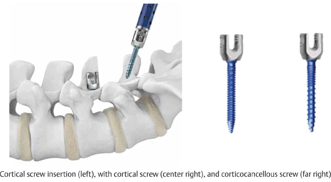
Table 6.2 DePuy Synthes Spine VIPER® Cortical Fix Screw System
Design | |
Set screw specifications Tulip design to reduce anatomical impact | |
| |
Modular aspects and variations | |
Screw diameters 5–9 mm (1-mm increments) | Screw lengths 30–55 mm (5-mm increments) |
Procedures | |
MIS TLIF, MIS MIDLF | |
Radiographs unavailable | |
Supplemental fixation systems | |
DePuy Synthes EXPEDIUM® Spine System, VIPER® System | |
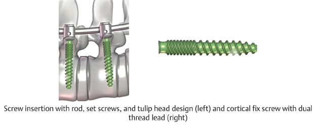
Table 6.3 Globus Medical CREO MCS™ Midline Cortical Stabilization System
Design | |
Set screw specifications Nonthreaded locking cap technology prevents cross-threading | Reduction capabilities 5 reduction options for up to 20 mm of reduction |
| |
Modular aspects and variations | |
Screw diameter 7 mm | Rod diameters 4.75, 5.5, 6.35 mm |
Procedures | |
MIS TLIF, MIS MIDLF | |
Radiographs unavailable | |
Supplemental fixation systems | |
Globus Medical REVOLVE® Posterior Stabilization System | |