Fig. 7.1
Schema of angular acceleration to the head
Angular acceleration produced by an impact can be applied to the head in three planes; sagittal, coronal and axial planes. In a practical sense, a pure impact involving only one of the three planes is feasible only in a controlled experimental situation. Angular acceleration in the coronal plane is most typically associated with diffuse brain injuries in primates (Gennarelli et al. 1982). On the other hand, acceleration forces (translational, rotational, or angular) in the sagittal plane may be associated with life-threating acute subdural hematoma: bleeding from disrupted bridging veins that course along the midline structures of the brain.
About 80 % of the knock-outs (KO) in professional boxing bouts involve concussions; hook shots to the head which result in an angular rotation of the neck produce the most severe type of KO (Tani et al. 2001).
Magnitude of acceleration (angular velocity and acceleration) is another important factor regarding concussion; more than 3,500 rad/s2 of angular acceleration is reported to produce a concussion (Shewchenko et al. 2005).
7.6 Experimental Studies of Concussion
In 1946 Pudenz and Shelden (1946) reported superficial brain deformation and shift occurred following a head impact to apes. Over thirty years later, Gennarelli et al. (1982) studied the mechanical and pathophysiological aspects of primate head injury. They reported that lateral rotational impacts to the head (coronal acceleration) were the most significant in the production of diffuse brain injury. Little academic research has been done on primates since Gennarelli’s report, although primate species present an obvious homology with humans.
7.7 Simulation of Concussions
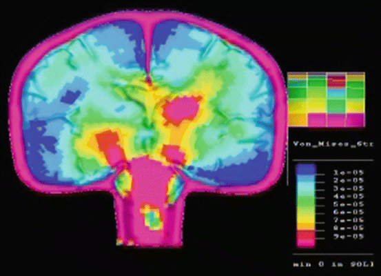
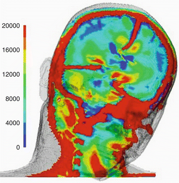
As primate based experiments have declined, computer-based physical simulation studies have simultaneously been increasing, both in frequency and in sophistication. The author and co-workers developed a 2D simulation study in which it was possible to demonstrate diffuse brain injury lesions similar to those seen in clinico-pathological settings. An impactor was applied to the right temporal side of the model at the level of the third ventricle, at a point which was at the same level as the center of gravity at a speed of 2 m/s. The model moved freely after the impact without any fixation. Major mechanical stresses on the brain parenchyma were observed in the midbrain, basal ganglia and corpus callosum underneath the falx cerebri (Fig. 7.2). Recent developments of the 3D voxel method enable it to provide a more precise representation of shear stress in the brain parenchyma after various impacts to the head in coronal and/or sagittal planes under different strain conditions (Fig. 7.3).


Fig. 7.2
2D simulation of diffuse brain injury

Fig. 7.3
3D voxel simulation of diffuse brain injury
Further development of the methods involved in simulation studies are expected to further elucidate the mechanical aspects of concussions.
7.8 Pathophysiology of Clinical Symptoms
Pathophysiological reactions and consequences of mechanical stress in brain parenchyma have been proposed as follows:
1.
Abrupt neuronal depolarization
2.
Release of excitatory neurotransmitters
3.
Ionic shifts
4.
Changes in glucose metabolism
5.
Altered cerebral blood flow
6.
Impaired axonal function
These pathological reactions may provoke various lesions and their associated loss of fiber connections in neuronal structures, and may result in transient brain dysfunction similar to that seen under various clinical syndromes. These pathological reactions are considered to be transient and reversible.
7.9 Aims of Prophylaxis for Concussions
Recent advances in sports medicine have reached the conclusion that concussions should be minimized. The main reason for prophylaxis of concussions is to avoid the following associated conditions:
1.


Second impact syndrome (SIS)
Stay updated, free articles. Join our Telegram channel

Full access? Get Clinical Tree


