Closed Reduction and Internal Fixation of Reverse Bennett’s Fractures

70 Closed Reduction and Internal Fixation of Reverse Bennett’s Fractures


Indications


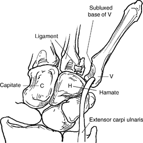
Base of the small metacarpal fracture with either or both displacement and joint subluxation. The entire ray is displaced with the radial intra-articular fragment of the metacarpal base remaining undisplaced on the hamate (H) (Fig. 70-1).


Technique


  • Regional anesthesia with intravenous sedation
  • Fracture reduction with longitudinal traction. Direct pressure to the small metacarpal base toward the avulsion fracture facilitates reduction
  • Main focus is to reduce metacarpal subluxation and restore articular congruity.
  • Reduction verified using fluoroscopy.
  • Percutaneous fixation with two oblique 0.45 in. (1.1 mm) trocar-tipped wires drilled through the small metacarpal. One pin is directed into the ring metacarpal and the other is placed in an oblique direction into the carpus (Fig. 70-2A-C).
  • Adjust pin position and length using fluoroscopy. The wires are either retained in a percutaneous position or preferably cut beneath the skin.

image

Only gold members can continue reading. Log In or Register to continue

Stay updated, free articles. Join our Telegram channel

Mar 21, 2017 | Posted by in MUSCULOSKELETAL MEDICINE | Comments Off on Closed Reduction and Internal Fixation of Reverse Bennett’s Fractures

Full access? Get Clinical Tree

Get Clinical Tree app for offline access