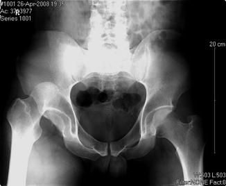
Fig. 11.1
Trauma patient. The figure depicts a trauma patient with multisystem pathology. Such patients can have numerous orthopaedic injuries and management of these injuries needs to be in accordance with damage control orthopaedic protocols
Damage control was a term originally coined by the United States navy to describe tactics needed to keep compromised vessels afloat. Originally adopted by Rontondo et al., “damage control surgery” was a method of treatment that used rapid but non-definitive control of hemorrhage to avoid the lethal triad of acidosis, hypothermia, and coagulopathy in patients exsanguinating from penetrating abdominal wounds [5]. This idea was later adapted to fit orthopaedic protocols of fixation. Damage Control Orthopaedics is an adaptive treatment strategy defined as the provisional stabilization of musculoskeletal injuries in order to allow the patient’s overall physiology to improve to tolerate longer more taxing procedures required for definitive fixation [2]. Under this definition, the primary tools for early stabilization were splinting, traction, and external fixation. With use of DCO, orthopaedic intervention in the acute injury phase is limited to temporary stabilization of structures that could contribute to physiological compromise. Too early an intervention could potentially add or trigger greater physiological injury, called the “2nd hit.”
The “2nd” hit concept describes the additive orthopaedic impact after the “1st hit” which is the initial trauma. The 1st hit can activate inflammatory mediators in some patients (interleukins) resulting in a hyperreactive physiology. The subsequent “2nd hit” can trigger an even greater response that can result in further end-organ injury and patient demise. Too early an orthopaedic intervention can be the cause of the 2nd hit via substantial blood loss and/or further soft-tissue damage. The end result may be hypoperfusion, hypoxia/ischemia, reperfusion, and tissue damage causing local necrosis, inflammation, and acidosis [2]. This inflammatory response to trauma, whether it be the “first” or “second” hit, that causes release of proinflammatory cytokines, proteins, and hormones was probably the cause of the major complications seen with aggressive, early fixation like ARDS and multiorgan failure [2]. DCO arose with a purpose of avoiding worsening of physiological parameters by delaying definitive fixation until the patient’s physiology permitted.
The decision to employ DCO can be decided upon using genetic and numerous biochemical markers, but these analyses are not available in a timely manner at most facilities so physiological signs can be used to guide decision-making [2]. Some particular physiological markers were defined by Pape et al. to be serum lactate greater than 2.5 (mmol/L), base excess of more than 8 (mmol/L), a pH of less than 7.24, and temperature less than 35 °C, surgical time more than 90 min, any coagulopathy, and transfusion of more than 10 units of packed red blood cells [6]. Some fractures should still be approached with caution even if all physiological parameters indicate lower risks for second hit phenomenon. Recognizing that the above parameters are proxies for complex interactions, there are undoubtedly physiological or genetic predispositions that remain unrecognized that could still result in complications in the absence of overtly abnormal markers. From an orthopaedic standpoint, femur fractures in polytrauma patients, pelvic ring injuries with substantial hemorrhage, and geriatric polytrauma patients are those at risk [2]. While definitive treatment will be covered in different chapters, the decision to undergo definitive surgery can be done when the criteria in Table 11.1 or when fluid balance is negative [2]. For this chapter, however, we will focus on noninvasive and invasive methods to initially stabilize fractures in trauma patients.
Table 11.1
Parameters to consider when deciding to implement damage control orthopaedic protocol
Polytrauma with Injury Severity Score of >20 points with additional thoracic trauma (Abbreviated Injury Scale score of >2 points) |
Polytrauma with abdominal and pelvic injuries and hemorrhagic shock (systolic blood pressure <90 mmHg) |
Injury Severity Score of ≥40 points without additional thoracic injury |
Initial pulmonary artery pressure of >24 mmHg |
Increased pulmonary artery pressure of >6 mmHg during intramedullary nailing |
Difficult resuscitation |
Platelet count <90,000/μ[micro]L (<90 × 109/L) |
Hypothermia (e.g., temperature of <35 °C) |
Transfusion of >10 units of blood |
Bilateral lung contusion on initial chest radiograph |
Multiple long-bone fractures and truncal injury |
Prolonged duration of anticipated surgery (>90 min) |
Assessment
Initial presentation of the trauma patient needs to be handled in a systematic way to ensure proper assessment and workup usually according to BLS/ACLS/ATLS protocols [7]. Regardless of the protocol chosen, the ABCs (airway, breathing, and circulation) need to be assessed first to ensure all vital systems are competent. Next, the rest of the patient is assessed with higher priority ailments receiving the most attention. The general rule in trauma is life over limb, but at times extremity injuries can be life threatening.
From the orthopaedic standpoint, a proper examination of the extremities includes inspecting for open wounds or soft-tissue defects and palpation for point tenderness or crepitus over all boney structures. Next, one should range all major joints paying particular attention to decreased or painful range of motion, indication of possible dislocation, with additional investigation for laxity or instability hinting at ligamentous injury. Pulses and neurological status need to be assessed to determine if any vascular or nerve damage is present in the limb. If such a deficit is found, need for further investigation is indicated and urgent. Such exams like ankle-brachial index or computed tomography angiography are used rule out vascular injury/compromise. Presence of neurological deficits, while usually not critical, can hint at a larger problem such as central neurological compromise or severe peripheral nerve damage that could affect future functioning of the limb. In patients with altered consciousness, only signs of grimacing or withdrawal may be the only indicators of injury. In obtunded or intubated patients, the clinical exam requires more attention as it is rather common to miss orthopaedic injuries in these patients.
Each limb needs to be assessed for compartment syndrome due to the devastating effects of a missed diagnosis that can ultimately end in loss of function or even amputation of a limb. Compartment syndrome is also a leading cause of litigation in orthopaedics. It is usually not what is done, but rather, what is not done that becomes the focus of litigation in such cases. Providers need to recognize that the presence of an open fracture does not exclude a compartment syndrome [8] since the small opening of the skin and fascia do not usually result in an adequate decompression, and there are other compartments remote from the open injury that can be involved. All details of compartment syndrome are discussed in more detail in Chap. 10.
Radiography
Once the full assessment is completed and fluid resuscitation according to ATLS has begun [8], x-rays of areas that are suspicious for fracture need to be evaluated radiographically. The basic tenets of appropriate radiographic evaluation are to obtain images of joints proximal and distal to observed fractures. This rule is important because forces in any given bone are by definition transmitted to the joint “above and below” and can result in remote injury. For example, axial loads from the foot can result in injuries to the hip or knee. The physics of load transfer helps explain a special situation that exists in the forearm and lower leg. The presence of the interosseous membrane in the forearm and lower leg, where it contributes to a structure called the syndesmosis, acts as a structure distributing forces around the region causing associated fractures and dislocations [9, 10] (see Fig. 11.2). This explains why severe external rotation about the ankle can cause fractures in the proximal lower leg about the knee or why a fracture of the radial or ulnar shafts can cause a secondary dislocation or fracture of the other bone (see Fig. 11.3).
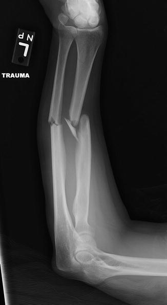
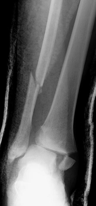
Fig. 11.2
Both-bone forearm fracture. An AP x-ray showing a both-bone forearm fracture with comminution of the radial shaft fracture
Fig. 11.3
Ankle fracture with associated syndesmosis injury. The figure shows a distal fibular fracture along with a medial malleolar fracture. A syndesmosis injury can also be noted secondary to the increased space between the fibula and tibia where overlap between the bones is usually expected
If pelvic fractures are suspected from the initial screening, AP pelvis film, Judet films, and inlet and outlet views should be taken. An AP film is a good screening tool that can direct the need for different sets of films (see Fig. 11.4). Judet films allow for the evaluation of posterior and anterior columns as well as the posterior and anterior acetabular walls. Inlet views of the pelvis allow for proper evaluation of any anterior/posterior displacement of the sacroiliac joint sacrum or iliac wing as well as determining rotation deformities of the ilium and sacral impaction injuries. Outlet views allow for evaluation of vertical displacement of the hemipelvis (see Fig. 11.5). However, with the implementation of new technology, CT scanning is generally the most informative and thus definitive radiographic technique. Newer CT radiographs may supplant the use of common x-rays, especially when done in a suboptimal manner.
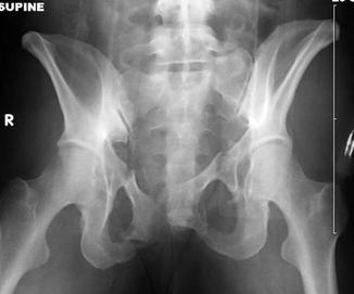
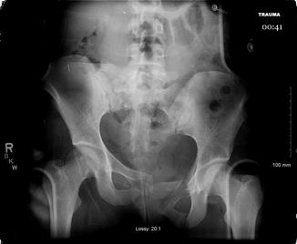
Fig. 11.4
Bilateral inferior/superior rami fractures. An AP film of the pelvis depicting a bilateral superior and inferior pubic rami fractures in a trauma patient (Photo courtesy of Evan Siegall, MD)
Fig. 11.5
Vertical sheer pelvis. This is an AP x-ray of the pelvis showing displacement of the left hemipelvis secondary to severe trauma. Such injuries are called vertical sheer injuries and are indicative of high-energy trauma
Noninvasive Techniques of Treatment
Closed fractures and dislocations can be dealt with numerous ways in trauma patients; the least traumatic techniques are noninvasive ones, which are reduction, traction, and splinting. When diagnosed clinically and confirmed radiographically, all dislocations need to be reduced as soon as possible. The need for immediate joint reduction is owed to the fact that joint dislocations can stretch/compromise nearby neurovascular structures. Neurological or vascular deficits may result as a result of prolonged dislocation that either compresses or stretches such structures. Specific examples of this can be seen in patients with dislocated hips. These patients are at an increased risk of osteonecrosis of the femoral head secondary to the hip capsule being stretched or torn which contains the vessels that supply the femoral head. Sciatic nerve injury can also occur the longer the femoral head stays dislocated and compressing the sciatic nerve (see Fig. 11.6). The effects of nerve damage secondary to dislocation are also seen in the shoulder where injury to the axillary nerve or brachial plexus can occur (see Fig. 11.7).