Clinodactyly
Robert Carrigan
DEFINITION
Clinodactyly refers to an abnormal about of radioulnar angulation of a digit (>15 degrees).
The small finger is most commonly observed.
This condition is often bilateral.
ANATOMY
The finger consists of three phalanges (proximal, middle, and distal).
The normal phalangeal physis is located at the proximal portion of each phalanx.
PATHOGENESIS
The angulation is result of abnormal development of one of the phalanges (most often the middle phalanx [p2]).
Abnormal development of the phalanx may be due to an irregular physis (longitudinal bracket epiphysis). This may also be referred to as a delta phalanx.
The tethering effect of the bracket epiphysis on the radial side of the finger causes abnormal growth of the phalanx resulting in a triangular or trapezoidal shape.
Extra bones may be encountered.
NATURAL HISTORY
The natural history of clinodactyly is variable and poorly documented, owing to the great number of cases that are asymptomatic and do not require treatment.
Angulation may be stable or rapidly progressive at times of growth, depending on the extent of the involvement of the physis and/or presence of extra phalanges.
PATIENT HISTORY AND PHYSICAL EXAM FINDINGS
Clinodactyly may be present at birth or develop during a period of growth (FIG 1).
Clinodactyly is often bilateral in the small finger.
Clinodactyly is an autosomal dominant condition with variable penetration.
Involvement of the thumb is rare and is associated with varying syndromes.
IMAGING AND OTHER DIAGNOSTIC STUDIES
Standard radiographs (three views: anteroposterior[AP], lateral [LAT], and oblique [OBL]) of the hand and affected digit are sufficient to determine the area of involvement.
Contralateral images are useful for comparison.
Advanced imaging such as computed tomography (CT) is rarely needed. Magnetic resonance imaging (MRI) may be useful to delineate the shape of a bracket diaphysis.
DIFFERENTIAL DIAGNOSIS
The diagnosis of clinodactyly is straightforward; clinical examination and radiographs are sufficient to make the diagnosis.
Associated syndrome should be screened for, these include Down, Rubinstein-Taybi, Apert, and Russell-Silver.
NONOPERATIVE MANAGEMENT
Observation may be considered for angulated digits that do not impair function. Splinting is not effective.
Most cases can be treated nonoperatively; surgery should be considered for significant angular deformity that compromises hand function.
SURGICAL MANAGEMENT
Preoperative Planning
Timing of surgery is variable, depending on the degree of angulation and how much growth potential remains.
Small amounts of angulation with little remaining growth potential may be addressed when the child is older.
Larger amounts of angulation or children with the potential for worsening angulation may consider earlier intervention.
Positioning
The patient is positioned supine on the operating room table and the body is pulled over to the affected side.
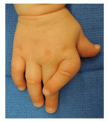
FIG 1 • Clinodactyly of the index finger from osteochondroma in a child with multiple hereditary exostosis.Related posts:
 Anatomy and Surgical Approaches of the Forearm, Wrist, and Hand
 Arthroscopic Treatment of Chondral Injuries and Osteochondritis Dissecans
 Fragment-Specific Fixation of Distal Radius Fractures
 Arthroscopic and Open Triangular Fibrocartilage Complex Repair
 Carpal Tunnel Release: Endoscopic, Open, and Revision
 Surgical Treatment of Acute and Chronic Paronychia and Felons

Stay updated, free articles. Join our Telegram channel

Full access? Get Clinical Tree

 Get Clinical Tree app for offline access
