

Fig. 1.1
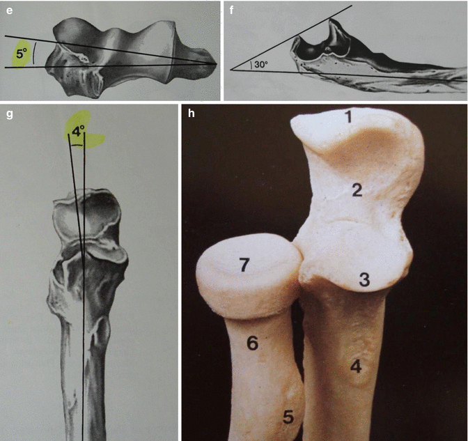
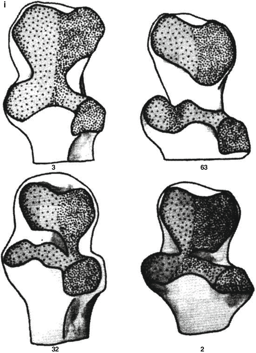
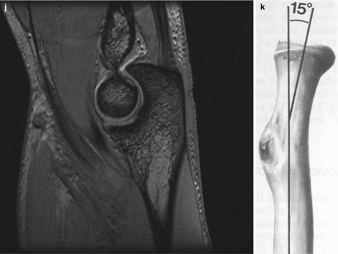
(a) Anterior view: lateral epicondyle (1), capitellum (2), trochlea (3), medial epicondyle (4), coronoid process (5), and radial head (6). (b) Posterior view: olecranon (7). (c–e) The condyles of the humerus show a 30° anterior flexion in relation to the long axis, a 6–8° valgus tilt, and a 5° internal rotation in relation to the epicondylar line. (f, g) In the proximal ulna, the trochlear notch forms an angle of 30° with the ulna shaft, and there is also a slight 4° valgus angulation of the ulnar shaft. (h–j) The hyaline cartilage distribution of the proximal ulna varies and is often misinterpreted as osteochondral damage. (k) The radial head forms a 15° angle with the axis of the radial shaft
The shaft of the humerus ends in a lateral and medial ridge. Approximately 12 cm above the lateral ridge is a sulcus in which the radial nerve passes to the lateral side of the humerus. This is an important anatomical landmark in the surgical treatment of humeral fractures with plates or external fixators. Lateral and medial ridges end distally in the lateral and medial epicondyles (see Fig. 1.1a, b). The condyles of the humerus show a 30° anterior flexion in relation to the long axis, a 6–8° valgus tilt, and a 5° internal rotation in relation to the epicondylar line (see Fig. 1.1c–e).
To prevent anterior impingement during flexion of the elbow, the coronoid fossa and the radial fossa are located between the lateral and medial ridges on the anterior side of the distal humerus. On the posterior side, the olecranon fossa is located between the epicondyles to prevent posterior impingement during extension.
The trochlea is formed by the medial epicondyle, which forms the ulnohumeral joint with the olecranon of the ulna, which stabilizes the elbow during extension. The anterior side of the lateral epicondyle forms the capitellum. This convex structure articulates with the concave surface of the radial head. This is the radiohumeral joint, which plays a role in the stability of the elbow in flexion.
In the proximal ulna, the trochlear notch forms an angle of 30° with the ulna shaft, and there is also a slight 4° valgus angulation of the ulnar shaft (see Fig. 1.1f, g). The trochlear notch is divided into an anterior and a posterior part by the incisura trochlearis, a transverse portion composed of fatty tissue. This area of the olecranon can be used during an olecranon osteotomy to minimize cartilage damage. The hyaline cartilage distribution of the proximal ulna varies and is often misinterpreted as osteochondral damage (see Fig. 1.1h–j). The coronoid process, a protuberance of the ulna that demarcates the trochlear notch anteriorly, often fractures during dislocation of the elbow. Just distal and radial to the coronoid process, the radial notch of the ulna articulates with the radial head in the proximal radioulnar joint, contributing to pronation and supination of the forearm.
Since the radial head articulates with both the capitellum of the humerus and the radial notch of the ulna, it is covered with cartilage 280° around. The uncovered part of the radial head can be used for screw fixation in case of radial head fractures. The radial head forms a 15° angle with the axis of the radial shaft (see Fig. 1.1k).
There is a great amount of congruency between the articulating surfaces of the elbow. The tongue and groove-like fitting of the distal humerus on the ulna and radius make medial and lateral gliding almost impossible [2, 3].
The articular contact is influenced by the position of the elbow and the forearm. The radial head makes no contact with the cartilage of the capitellum during extension of the elbow. However, during flexion the radial head moves proximally resulting in an increased contact with the distal humerus. Supination of the forearm decreases the radiocapitellar contact, while pronation increases it. The knowledge of these positions is important during clinical examination of a degenerative elbow [4].
1.2 Joint Capsule and Ligaments
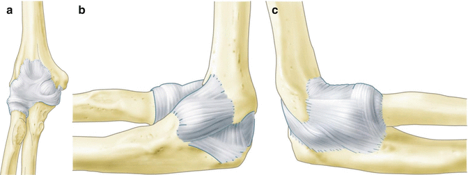
The three elbow joints are surrounded by a joint capsule. This capsule includes the olecranon, the coronoid fossa, and the radial fossa but not the humeral epicondyles. At the level of the radial head, distal from the radial annular ligament, the joint capsule forms a recess to preserve a good rotation of the radius (see Fig. 1.2a).


Fig. 1.2
(a) Anterior view of the joint capsule of the elbow. (b) Medial collateral ligement consisting of an anterior (AMCL) and posterior (PMCL) bundle and a transversal ligament. (c) Lateral collateral ligament complex consisting of the lateral ulnar collateral ligament (LUCL), the radial collateral ligament (RCL) and the annular ligament (AL)
The joint capsule has a limited role in the stability of the elbow. To allow flexion and extension of the elbow, the capsule is loose on the anterior side and especially on the posterior. The volume of the capsule has been shown to average 23 ml. The capsule is most lax at 80° of flexion. Therefore patients with acute joint injury and inflammation combined with joint effusion find this position more comfortable. To prevent the capsule from sticking into the joints, small articular muscles radiate from the triceps brachii muscle and the brachial muscle. These muscles maintain sufficient tension on the capsule [5].
The collateral ligaments of the elbow are formed by thickenings of the capsule on the medial and lateral side. The medial collateral ligament consists of an anterior (AMCL) and a posterior (PMCL) bundle and a transversal ligament (also known as the Cooper ligament). The anterior and posterior bundles originate from the medial humeral epicondyle. The anterior bundle inserts the base of the coronoid process (sublime tubercle) of the ulna, and the posterior bundle inserts the medial part of the olecranon. The mean length of the AMCL is 27.1 mm and that of PMCL is 24.2 mm; the mean widths are about 4.7 mm and 5.3 mm, respectively. The function of these ligaments is to restrain valgus stress during extension (anterior bundle) and during flexion (posterior bundle) (see Fig. 1.2b) [6]. Studies reveal that the AMCL can be subdivided into three regions or bands according to their function [7, 8].
Stay updated, free articles. Join our Telegram channel

Full access? Get Clinical Tree


