Fig. 4.1
Prevalence of cardiovascular disease by age and sex in adults age 20 and older: NHANES 2003–2006 (Source: NCHS and NHLBI. These data include CHD, HF, stroke, and hypertension reproduced with permission from AHA Statistical Update [1])
Atherosclerosis is a progressive disease of the intima and sub-intimal layers of blood vessels with each stage marked by increasing amounts of inflammatory mediators, smooth muscle cells, connective tissue and lipids resulting in an overall increase in atheroma size relative to the blood vessel lumen (Fig. 4.2) [2].
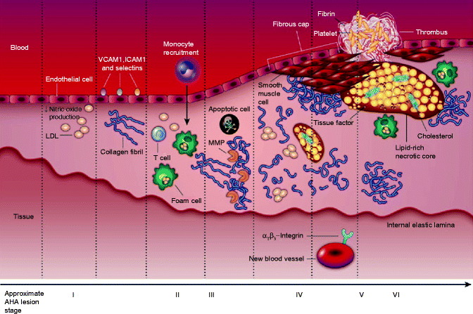

Fig. 4.2
The progression of an atherosclerotic plaque, from a normal blood vessel (far left) to a vessel with a ruptured atherosclerotic plaque with thrombus formation (far right); ICAM1 intercellular adhesion molecule 1, LDL low density lipoprotein, MMP matrix metalloproteinase, VCAM1 vascular cell-adhesion molecule 1 (Reproduced with permission of Macmillan Publishers Ltd from Sanz and Fayad [2])
The clinical manifestations of atherosclerosis vary from the asymptomatic state in the early stage of atherosclerosis to transient, symptomatic ischemia secondary to supply demand mismatch from increasing plaque formation seen in later stages.
These stable clinical symptoms depend upon the vascular territory involved but most commonly involve the cardiovascular, cerebrovascular, and peripheral vascular beds (Fig. 4.3) [3].
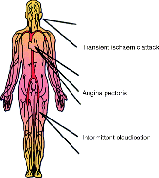

Fig. 4.3
Major clinical manifestations of atherosclerotic disease (Reproduced with permission of Oxford University Press from Viles-Gonzalez et al. [3])
The process of atherothrombosis involves the acute formation of a platelet rich thrombus on the surface of an unstable, eroded, or ruptured atherosclerotic plaque (Fig. 4.4) [4].
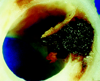

Fig. 4.4
Macroscopic view of a ruptured coronary plaque (Reproduced with permission of BMJ Group from Davies [4])
Due to the abrupt nature of atherothrombotic events, the clinical manifestations are generally more acute, necessitating prompt diagnosis and intervention.
The three most common clinical manifestations of acute atherothrombosis include acute coronary syndrome, stroke (cerebrovascular accident), and intermittent claudication with critical limb ischemia of the peripheral arterial tree.
Coronary Artery Atherothrombosis
Clinically stable atherosclerosis of the coronary arterial bed is generally asymptomatic or manifests as stable angina, the latter of which increases in prevalence with increasing age in both men and women (Table 4.1) [5].
Table 4.1
Prevalence of angina pectoris by age and sexa
Percent of population
Age
Men
Women
55–64
7.6
3.4b
65–74
18.2
7.7
75–84
23.1
17.2
85–94
30.2
14.4
As the atherosclerotic plaque enlarges, in most cases the initial compensation is an overall enlargement of the vessel wall area with overall preservation of the arterial lumen (positive remodeling) (Figs. 4.5 and 4.6) [6].
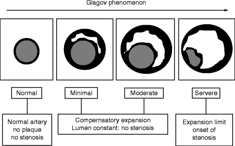
Fig. 4.5
Cross sectional view of the arterial lumen demonstrating the initial preservation of the arterial lumen in the early stages of atherosclerotic accumulation
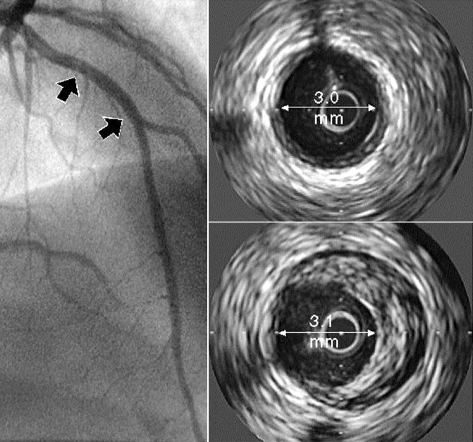
Fig. 4.6
“Normal” coronary angiography of the left anterior descending artery (left) associated with positive remodeling and a large, eccentric plaque on intravascular ultrasound (lower right), (arrow) lumen diameter (Reproduced with permission from Nissen and Yock [6])
Symptoms of stable angina evolve as the positive remodeling process is exhausted and the atherosclerotic plaque encroaches on the coronary vessel lumen (negative remodeling) reducing coronary flow reserve (generally 50 % diameter stenosis).
The reduction in coronary flow reserve leads to a reduction in myocardial oxygen supply during times of increased metabolic demand (i.e., exercise) (Fig. 4.7) [7].
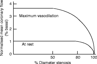
Fig. 4.7
The relationship between resting and maximal coronary blood flow as a function of percent diameter stenosis (Reproduced with permission of Elsevier from Gould et al. [7])
Stable angina is well-described clinically and classically involves a syndrome of stress related substernal chest pressure with or without associated shortness of breath, nausea/vomiting, and radiation down the left arm or up into the neck or jaw that resolves completely with rest or nitroglycerine.
The pretest probability of chest pain symptoms resulting from obstructive coronary disease can be estimated based upon age, gender, and how many typical angina features (substernal location, related to stress/exertion, alleviated with rest or nitroglycerine) (Table 4.2) [8].
Table 4.2
Pretest probability of coronary artery disease by age, gender, and symptoms
Age (years)
Gender
Typical/definite angina pectoris
Atypical/probable angina pectoris
Nonanginal chest pain
Asymptomatic
30–39
Men
Intermediate
Intermediate
Low
Very low
Women
Intermediate
Very low
Very low
Very low
40–49
Men
High
Intermediate
Intermediate
Low
Women
Intermediate
Low
Very low
Very low
50–59
Men
High
Intermediate
Intermediate
Low
Women
Intermediate
Intermediate
Low
Very low
60–69
Men
High
Intermediate
Intermediate
Low
Women
High
Intermediate
Intermediate
Low
The most common cause of acute coronary atherothrombosis is plaque rupture followed by plaque erosion and, infrequently, a calcified nodule [9].
The precursor to plaque rupture is postulated to be the thin cap fibroatheroma (TCFA) which consists of a necrotic, inflammatory core that is separated by the arterial lumen by a thin, fibrous cap (Fig. 4.8) [9, 10].
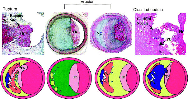
Fig. 4.8
Atherosclerotic lesions with luminal thrombi. Ruptured plaques are thin fibrous cap atheromas (thin, fibrous cap <65 μm) with luminal thrombi (Th). The thrombus is in communication with the lipid rich necrotic core (NC). Erosions occur over lesions rich in smooth muscle and do not, commonly, have a necrotic core. Calcified nodules are plaques with luminal thrombi showing calcific nodules protruding into the lumen through a disrupted fibrous cap (FC) (Reproduced with permission from Virmani et al. [10])
The TCFA is most frequently observed in patients dying from an acute myocardial infarction, has been found most frequently in proximal coronary arteries, and has been found associated with positive remodeling and less than 50 % diameter stenoses [9].
Plaque erosion is defined as an acute thrombus in direct contact with the intima, in an area of absent endothelium. They tend to be infrequently calcified and associated with a thick fibrous cap with no established communication with the deep, underlying plaque (Fig. 4.8) [10].
Plaque erosion has been shown to occur more frequently in younger patients, particularly in women less than 50 years of age [9].
Calcified nodules refer to lesions with fibrous cap disruption and thrombi associated with eruptive, dense, calcific nodules (Fig. 4.8) [10].
Calcified nodules appear to be associated with healed plaques and are found predominantly in the mid right coronary artery [10].
Plaque rupture and erosion result in platelet activation and ultimately the formation of a platelet-rich thrombus, an unstable event which has a spectrum of clinical presentations ranging from ST segment elevation acute coronary syndrome (STEMI) to non-ST segment elevation acute coronary syndrome (comprising unstable angina and non-ST segment elevation myocardial infarction [NSTEMI]).
The most dramatic presentation of acute coronary atherothrombosis is seen clinically as an ST segment elevation acute myocardial infarction (STEMI) which is generally associated with a complete occlusion of a coronary artery and represents a medical emergency (Fig. 4.9).
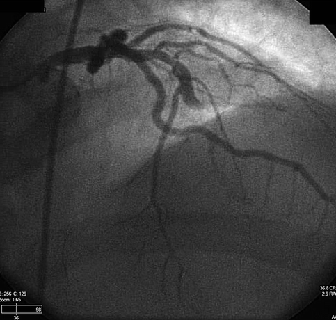
Fig. 4.9
Anterior-posterior view with cranial angulation of the left coronary tree showing a complete occlusion of the mid left anterior descending artery
Patients typically present with the acute onset of anginal symptoms at rest with or without associated shortness of breath, nausea, vomiting, and diaphoresis.
Physical examination can range from normal to overt cardiogenic shock with pulmonary edema from acute left ventricular failure.
The electrocardiogram is notable for ST segment elevation in leads associated with the vascular territory involved (Fig. 4.10).
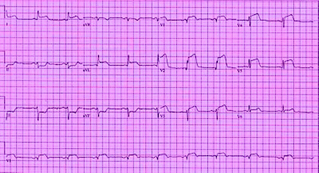
Fig. 4.10
An anterior-lateral STEMI with ST elevation in leads V1–V6, I, aVL associated with reciprocal changes in leads II, III, and aVF
The acute atherothrombotic event resulting in NSTE-ACS can translate clinically as new onset, severe angina, an increase in the frequency and/or intensity of current anginal symptoms, or as rest angina.
Stay updated, free articles. Join our Telegram channel

Full access? Get Clinical Tree


